 Movie
Movie Controller


+ Open data

- Basic information
| Entry | Database: PDB / ID: 4oyn | ||||||
|---|---|---|---|---|---|---|---|
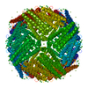
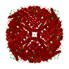
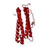
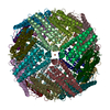
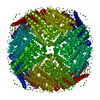
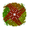
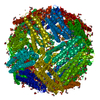
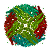
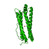
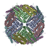
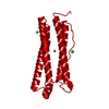
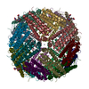
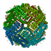
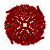
| Title | Fifteen minutes iron loaded human H ferritin | ||||||
 Components Components | Ferritin heavy chain | ||||||
 Keywords Keywords | OXIDOREDUCTASE / ferroxidase / iron loaded / human H ferritin / iron storage protein | ||||||
| Function / homology |  Function and homology information Function and homology informationiron ion sequestering activity / ferritin complex / Scavenging by Class A Receptors / Golgi Associated Vesicle Biogenesis / ferroxidase / negative regulation of ferroptosis / autolysosome / ferroxidase activity / negative regulation of fibroblast proliferation / ferric iron binding ...iron ion sequestering activity / ferritin complex / Scavenging by Class A Receptors / Golgi Associated Vesicle Biogenesis / ferroxidase / negative regulation of ferroptosis / autolysosome / ferroxidase activity / negative regulation of fibroblast proliferation / ferric iron binding / autophagosome / iron ion transport / ferrous iron binding / Iron uptake and transport / tertiary granule lumen / ficolin-1-rich granule lumen / intracellular iron ion homeostasis / immune response / iron ion binding / negative regulation of cell population proliferation / Neutrophil degranulation / extracellular exosome / extracellular region / identical protein binding / nucleus / cytoplasm / cytosol Similarity search - Function | ||||||
| Biological species |  Homo sapiens (human) Homo sapiens (human) | ||||||
| Method |  X-RAY DIFFRACTION / SYNCHROTRON / MOLECULAR REPLACEMENT / Resolution: 1.43 Å X-RAY DIFFRACTION / SYNCHROTRON / MOLECULAR REPLACEMENT / Resolution: 1.43 Å | ||||||
 Authors Authors | Pozzi, C. / Di Pisa, F. / Mangani, S. | ||||||
 Citation Citation | Journal: Acta Crystallogr.,Sect.D / Year: 2015 Title: Iron binding to human heavy-chain ferritin. Authors: Pozzi, C. / Di Pisa, F. / Bernacchioni, C. / Ciambellotti, S. / Turano, P. / Mangani, S. | ||||||
| History |
|
- Structure visualization
| Structure viewer | Molecule: MolmilJmol/JSmol |
|---|
- Downloads & links
-Download
| PDBx/mmCIF format | 4oyn.cif.gz | 66.2 KB | Display | PDBx/mmCIF format |
|---|---|---|---|---|
| PDB format | pdb4oyn.ent.gz | 47 KB | Display | PDB format |
| PDBx/mmJSON format | 4oyn.json.gz | Tree view | PDBx/mmJSON format | |
| Others |  Other downloads Other downloads |
-Validation report
| Arichive directory | https://data.pdbj.org/pub/pdb/validation_reports/oy/4oynftp://data.pdbj.org/pub/pdb/validation_reports/oy/4oyn | HTTPS FTP |
|---|
-Related structure data
| Related structure data |  4y08C  4ykhC  4zjkC  3ajoS  4p03 S: Starting model for refinement C: citing same article ( |
|---|---|
| Similar structure data |


-Links
 PDBj
PDBj


- Assembly
| Deposited unit | 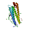
| ||||||||||||||||||||||||||||||||||||
|---|---|---|---|---|---|---|---|---|---|---|---|---|---|---|---|---|---|---|---|---|---|---|---|---|---|---|---|---|---|---|---|---|---|---|---|---|---|
| 1 | x 24
| ||||||||||||||||||||||||||||||||||||
| Unit cell |
| ||||||||||||||||||||||||||||||||||||
| Components on special symmetry positions |
|
-Components
-Protein , 1 types, 1 molecules A
| #1: Protein | Mass: 21255.656 Da / Num. of mol.: 1 Source method: isolated from a genetically manipulated source Source: (gene. exp.) Homo sapiens (human) / Gene: FTH1, FTH, FTHL6, OK/SW-cl.84, PIG15 / Production host:  |
|---|
-Non-polymers , 6 types, 345 molecules 


| #2: Chemical | ChemComp-TRS /  Mass: 122.143 Da / Num. of mol.: 1 / Source method: obtained synthetically / Formula: C4H12NO3 / Comment: pH buffer*YM Mass: 122.143 Da / Num. of mol.: 1 / Source method: obtained synthetically / Formula: C4H12NO3 / Comment: pH buffer*YM | ||||||
|---|---|---|---|---|---|---|---|
| #3: Chemical | ChemComp-BCN /  Mass: 163.172 Da / Num. of mol.: 1 / Source method: obtained synthetically / Formula: C6H13NO4 / Comment: pH buffer*YM Mass: 163.172 Da / Num. of mol.: 1 / Source method: obtained synthetically / Formula: C6H13NO4 / Comment: pH buffer*YM | ||||||
| #4: Chemical | ChemComp-FE /  Mass: 55.845 Da / Num. of mol.: 7 / Source method: obtained synthetically / Formula: Fe Mass: 55.845 Da / Num. of mol.: 7 / Source method: obtained synthetically / Formula: Fe#5: Chemical | ChemComp-CL /  Mass: 35.453 Da / Num. of mol.: 13 / Source method: obtained synthetically / Formula: Cl Mass: 35.453 Da / Num. of mol.: 13 / Source method: obtained synthetically / Formula: Cl#6: Chemical |  Mass: 24.305 Da / Num. of mol.: 3 / Source method: obtained synthetically / Formula: Mg Mass: 24.305 Da / Num. of mol.: 3 / Source method: obtained synthetically / Formula: Mg#7: Water | ChemComp-HOH / | Mass: 18.015 Da / Num. of mol.: 320 / Source method: isolated from a natural source / Formula: H2O |
-Experimental details
-Experiment
| Experiment | Method: X-RAY DIFFRACTION |
|---|
- Sample preparation
| Crystal | Density Matthews: 3.15 Å3/Da / Density % sol: 60.9 % / Description: cubic crystals |
|---|---|
| Crystal grow | Temperature: 277 K / Method: vapor diffusion, hanging drop / pH: 9 / Details: 1.6 M MgCl2 and 0.1 M bicine pH 9.0 / PH range: 8 - 9 |
-Data collection
| Diffraction | Mean temperature: 100 K |
|---|---|
| Diffraction source | Source: SYNCHROTRON / Site: Diamond  / Beamline: I04-1 / Wavelength: 0.92 Å / Beamline: I04-1 / Wavelength: 0.92 Å |
| Detector | Type: DECTRIS PILATUS 2M / Detector: PIXEL / Date: Jul 5, 2013 |
| Radiation | Protocol: SINGLE WAVELENGTH / Monochromatic (M) / Laue (L): M / Scattering type: x-ray |
| Radiation wavelength | Wavelength: 0.92 Å / Relative weight: 1 |
| Reflection | Resolution: 1.43→32.51 Å / Num. obs: 49578 / % possible obs: 100 % / Observed criterion σ(I): 2 / Redundancy: 20 % / Rmerge(I) obs: 0.068 / Net I/σ(I): 27.9 |
| Reflection shell | Resolution: 1.43→1.51 Å / Redundancy: 20.1 % / Rmerge(I) obs: 0.417 / Mean I/σ(I) obs: 7.1 / % possible all: 100 |
- Processing
| Software | Name: REFMAC / Version: 5.7.0032 / Classification: refinement | ||||||||||||||||||||||||||||||||||||||||||||||||||||||||||||||||||||||||||||||||||||||||||||||||||||||||||||||||||||||||||||||||||||||||||||||||||||||||||||||||||||||||||||||||||||||
|---|---|---|---|---|---|---|---|---|---|---|---|---|---|---|---|---|---|---|---|---|---|---|---|---|---|---|---|---|---|---|---|---|---|---|---|---|---|---|---|---|---|---|---|---|---|---|---|---|---|---|---|---|---|---|---|---|---|---|---|---|---|---|---|---|---|---|---|---|---|---|---|---|---|---|---|---|---|---|---|---|---|---|---|---|---|---|---|---|---|---|---|---|---|---|---|---|---|---|---|---|---|---|---|---|---|---|---|---|---|---|---|---|---|---|---|---|---|---|---|---|---|---|---|---|---|---|---|---|---|---|---|---|---|---|---|---|---|---|---|---|---|---|---|---|---|---|---|---|---|---|---|---|---|---|---|---|---|---|---|---|---|---|---|---|---|---|---|---|---|---|---|---|---|---|---|---|---|---|---|---|---|---|---|
| Refinement | Method to determine structure: MOLECULAR REPLACEMENT Starting model: 3AJO Resolution: 1.43→31.09 Å / Cor.coef. Fo:Fc: 0.964 / Cor.coef. Fo:Fc free: 0.961 / SU B: 0.73 / SU ML: 0.029 / Cross valid method: FREE R-VALUE / ESU R: 0.053 / ESU R Free: 0.052 / Stereochemistry target values: MAXIMUM LIKELIHOOD
| ||||||||||||||||||||||||||||||||||||||||||||||||||||||||||||||||||||||||||||||||||||||||||||||||||||||||||||||||||||||||||||||||||||||||||||||||||||||||||||||||||||||||||||||||||||||
| Solvent computation | Ion probe radii: 0.8 Å / Shrinkage radii: 0.8 Å / VDW probe radii: 1.2 Å / Solvent model: MASK | ||||||||||||||||||||||||||||||||||||||||||||||||||||||||||||||||||||||||||||||||||||||||||||||||||||||||||||||||||||||||||||||||||||||||||||||||||||||||||||||||||||||||||||||||||||||
| Displacement parameters | Biso mean: 13.771 Å2
| ||||||||||||||||||||||||||||||||||||||||||||||||||||||||||||||||||||||||||||||||||||||||||||||||||||||||||||||||||||||||||||||||||||||||||||||||||||||||||||||||||||||||||||||||||||||
| Refinement step | Cycle: 1 / Resolution: 1.43→31.09 Å
| ||||||||||||||||||||||||||||||||||||||||||||||||||||||||||||||||||||||||||||||||||||||||||||||||||||||||||||||||||||||||||||||||||||||||||||||||||||||||||||||||||||||||||||||||||||||
| Refine LS restraints |
|