[English] 日本語
Yorodumi
- PDB-4ot4: X-ray Structure of the Adduct formed between cisplatin and Ribonu... -

+
Open data


ID or keywords:

Loading...

-
Basic information

Entry
Database: PDB / ID: 4ot4
TitleX-ray Structure of the Adduct formed between cisplatin and Ribonuclease A
ComponentsRibonuclease pancreaticPancreatic ribonuclease family
KeywordsHYDROLASE / RNase A fold
Function / homology
Function and homology information


pancreatic ribonuclease / ribonuclease A activity / RNA nuclease activity / nucleic acid binding / lyase activity / defense response to Gram-positive bacterium / extracellular region
Similarity search - Function
P-30 Protein / Ribonuclease A-like domain / Pancreatic ribonuclease / Ribonuclease A, active site / Ribonuclease A-domain / Ribonuclease A-like domain superfamily / Pancreatic ribonuclease / Pancreatic ribonuclease family signature. / Pancreatic ribonuclease / Roll / Alpha Beta
Similarity search - Domain/homology
Cisplatin / Ribonuclease pancreatic
Similarity search - Component
Biological speciesBos taurus (cattle)
MethodX-RAY DIFFRACTION / MOLECULAR REPLACEMENT / Resolution: 1.85 Å
AuthorsMessori, L. / Merlino, A.
CitationJournal: Inorg.Chem. / Year: 2014
Title: Cisplatin binding to proteins: molecular structure of the ribonuclease a adduct.
Authors: Messori, L. / Merlino, A.
History
DepositionFeb 13, 2014Deposition site: RCSB / Processing site: RCSB
Revision 1.0Jul 9, 2014Provider: repository / Type: Initial release
Revision 1.1Jul 16, 2014Group: Derived calculations
Revision 1.2Sep 20, 2023Group: Data collection / Database references ...Data collection / Database references / Derived calculations / Refinement description
Category: chem_comp_atom / chem_comp_bond ...chem_comp_atom / chem_comp_bond / database_2 / pdbx_initial_refinement_model / pdbx_struct_conn_angle / struct_conn / struct_site
Item: _database_2.pdbx_DOI / _database_2.pdbx_database_accession ..._database_2.pdbx_DOI / _database_2.pdbx_database_accession / _pdbx_struct_conn_angle.ptnr1_auth_comp_id / _pdbx_struct_conn_angle.ptnr1_auth_seq_id / _pdbx_struct_conn_angle.ptnr1_label_alt_id / _pdbx_struct_conn_angle.ptnr1_label_asym_id / _pdbx_struct_conn_angle.ptnr1_label_atom_id / _pdbx_struct_conn_angle.ptnr1_label_comp_id / _pdbx_struct_conn_angle.ptnr1_label_seq_id / _pdbx_struct_conn_angle.ptnr2_auth_seq_id / _pdbx_struct_conn_angle.ptnr2_label_alt_id / _pdbx_struct_conn_angle.ptnr2_label_asym_id / _pdbx_struct_conn_angle.ptnr3_auth_comp_id / _pdbx_struct_conn_angle.ptnr3_auth_seq_id / _pdbx_struct_conn_angle.ptnr3_label_alt_id / _pdbx_struct_conn_angle.ptnr3_label_asym_id / _pdbx_struct_conn_angle.ptnr3_label_atom_id / _pdbx_struct_conn_angle.ptnr3_label_comp_id / _pdbx_struct_conn_angle.ptnr3_label_seq_id / _pdbx_struct_conn_angle.value / _struct_conn.pdbx_dist_value / _struct_conn.pdbx_ptnr1_label_alt_id / _struct_conn.pdbx_ptnr2_label_alt_id / _struct_conn.ptnr1_auth_asym_id / _struct_conn.ptnr1_auth_comp_id / _struct_conn.ptnr1_auth_seq_id / _struct_conn.ptnr1_label_asym_id / _struct_conn.ptnr1_label_atom_id / _struct_conn.ptnr1_label_comp_id / _struct_conn.ptnr1_label_seq_id / _struct_conn.ptnr2_auth_asym_id / _struct_conn.ptnr2_auth_comp_id / _struct_conn.ptnr2_auth_seq_id / _struct_conn.ptnr2_label_asym_id / _struct_conn.ptnr2_label_atom_id / _struct_conn.ptnr2_label_comp_id / _struct_site.pdbx_auth_asym_id / _struct_site.pdbx_auth_comp_id / _struct_site.pdbx_auth_seq_id

-
Structure visualization

Structure viewerMolecule:
MolmilJmol/JSmol

Downloads & links

-
Assembly

Deposited unit
A: Ribonuclease pancreatic
B: Ribonuclease pancreatic
hetero molecules


Theoretical massNumber of molelcules
Total (without water)28,3175
Polymers27,4172
Non-polymers9003
Water2,252125
1
A: Ribonuclease pancreatic
hetero molecules


Theoretical massNumber of molelcules
Total (without water)14,3083
Polymers13,7081
Non-polymers6002
Water181
TypeNameSymmetry operationNumber
identity operation1_555x,y,z1
MethodPISA
2
B: Ribonuclease pancreatic
hetero molecules


Theoretical massNumber of molelcules
Total (without water)14,0082
Polymers13,7081
Non-polymers3001
Water181
TypeNameSymmetry operationNumber
identity operation1_555x,y,z1
MethodPISA
Unit cell
Length a, b, c (Å)99.767, 32.463, 67.575
Angle α, β, γ (deg.)90.00, 93.07, 90.00
Int Tables number5
Space group name H-MC121

-
Components

#1: Protein Ribonuclease pancreatic / Pancreatic ribonuclease family / RNase 1 / RNase A


Mass: 13708.326 Da / Num. of mol.: 2
Source method: isolated from a genetically manipulated source
Source: (gene. exp.) Bos taurus (cattle) / Gene: RNASE1, RNS1 / References: UniProt: P61823, EC: 3.1.27.5
#2: Chemical ChemComp-CPT / Cisplatin / diammine(dichloro)platinum / Cisplatin


Mass: 300.045 Da / Num. of mol.: 3 / Source method: obtained synthetically / Formula: Cl2H6N2Pt / Comment: medication, chemotherapy*YM
#3: Water ChemComp-HOH / water / Water


Mass: 18.015 Da / Num. of mol.: 125 / Source method: isolated from a natural source / Formula: H2O

-
Experimental details

-
Experiment

Experiment
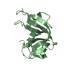
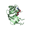
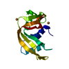
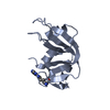
Method: X-RAY DIFFRACTION / Number of used crystals: 1

-
Sample preparation

CrystalDensity Matthews: 1.99 Å3/Da / Density % sol: 38.28 %
Crystal grow
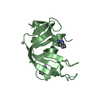
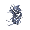
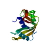
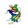
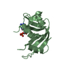
Temperature: 298 K / Method: vapor diffusion, hanging drop / pH: 5
Details: Crystals of RNase A were grown by hanging-drop vapor diffusion technique at 298 K mixing a protein solution containing 20 mg mL-1 of RNase A with equal volumes of reservoir solution. Well ...Details: Crystals of RNase A were grown by hanging-drop vapor diffusion technique at 298 K mixing a protein solution containing 20 mg mL-1 of RNase A with equal volumes of reservoir solution. Well diffracting crystals appeared within 15days from the following conditions:20% PEG4000 and 20 mM sodium citrate buffer pH 5.0. , VAPOR DIFFUSION, HANGING DROP

-
Data collection

DiffractionMean temperature: 100 K
Diffraction sourceSource: ROTATING ANODE / Type: RIGAKU MICROMAX-007 HF / Wavelength: 1.5418 Å
DetectorType: RIGAKU SATURN 944+ / Detector: CCD
RadiationMonochromator: graphite / Protocol: SINGLE WAVELENGTH / Monochromatic (M) / Laue (L): M / Scattering type: x-ray
Radiation wavelengthWavelength: 1.5418 Å / Relative weight: 1
ReflectionResolution: 1.85→50 Å / Num. all: 17075 / Num. obs: 17075 / Observed criterion σ(F): 0 / Observed criterion σ(I): 0

-
Processing

Software
NameVersionClassification
CrystalCleardata collection
PHASERphasing
REFMAC5.7.0032refinement
HKL-2000data reduction
HKL-2000data scaling
RefinementMethod to determine structure: MOLECULAR REPLACEMENT
Starting model: PDB code 1JVT
Resolution: 1.85→30.88 Å / Cor.coef. Fo:Fc: 0.952 / Cor.coef. Fo:Fc free: 0.915 / SU B: 3.675 / SU ML: 0.112 / Cross valid method: THROUGHOUT / σ(I): 0 / ESU R: 0.188 / ESU R Free: 0.172 / Stereochemistry target values: MAXIMUM LIKELIHOOD
Details: PHI/PSI TORSION ANGLES OUTSIDE THE EXPECTED RAMACHANDRAN PLOT ARE ASSOCIATED WITH RESIDUES THAT ARE NOT WELL DEFINED IN THE ELECTRON DENSITY MAPS (RESIDUES 16-22 OF THE TWO CHAINS AND ...Details: PHI/PSI TORSION ANGLES OUTSIDE THE EXPECTED RAMACHANDRAN PLOT ARE ASSOCIATED WITH RESIDUES THAT ARE NOT WELL DEFINED IN THE ELECTRON DENSITY MAPS (RESIDUES 16-22 OF THE TWO CHAINS AND RESIDUES IN CLOSE CONTACT WITH THESE REGIONS).
RfactorNum. reflection% reflectionSelection details
Rfree0.24709 880 5.2 %RANDOM
Rwork0.18924 ---
all0.192 16195 --
obs0.19235 16195 91.12 %-
Solvent computationIon probe radii: 0.8 Å / Shrinkage radii: 0.8 Å / VDW probe radii: 1.2 Å / Solvent model: MASK
Displacement parametersBiso mean: 27.794 Å2
Baniso -1Baniso -2Baniso -3
1-0.03 Å20 Å20.24 Å2
2--0.82 Å20 Å2
3----0.85 Å2
Refinement stepCycle: LAST / Resolution: 1.85→30.88 Å
ProteinNucleic acidLigandSolventTotal
Num. atoms1851 0 8 125 1984
Refine LS restraints
Refine-IDTypeDev idealDev ideal targetNumber
X-RAY DIFFRACTIONr_bond_refined_d0.0180.0191969
X-RAY DIFFRACTIONr_bond_other_d0.0020.021794
X-RAY DIFFRACTIONr_angle_refined_deg1.7991.9242670
X-RAY DIFFRACTIONr_angle_other_deg0.9133.0084132
X-RAY DIFFRACTIONr_dihedral_angle_1_deg8.5825253
X-RAY DIFFRACTIONr_dihedral_angle_2_deg35.30925.22788
X-RAY DIFFRACTIONr_dihedral_angle_3_deg13.06415339
X-RAY DIFFRACTIONr_dihedral_angle_4_deg17.15158
X-RAY DIFFRACTIONr_chiral_restr0.1150.2295
X-RAY DIFFRACTIONr_gen_planes_refined0.0090.022297
X-RAY DIFFRACTIONr_gen_planes_other0.0010.02455
X-RAY DIFFRACTIONr_nbd_refined
X-RAY DIFFRACTIONr_nbd_other
X-RAY DIFFRACTIONr_nbtor_refined
X-RAY DIFFRACTIONr_nbtor_other
X-RAY DIFFRACTIONr_xyhbond_nbd_refined
X-RAY DIFFRACTIONr_xyhbond_nbd_other
X-RAY DIFFRACTIONr_metal_ion_refined
X-RAY DIFFRACTIONr_metal_ion_other
X-RAY DIFFRACTIONr_symmetry_vdw_refined
X-RAY DIFFRACTIONr_symmetry_vdw_other
X-RAY DIFFRACTIONr_symmetry_hbond_refined
X-RAY DIFFRACTIONr_symmetry_hbond_other
X-RAY DIFFRACTIONr_symmetry_metal_ion_refined
X-RAY DIFFRACTIONr_symmetry_metal_ion_other
X-RAY DIFFRACTIONr_mcbond_it2.8132.7581009
X-RAY DIFFRACTIONr_mcbond_other2.8142.7581008
X-RAY DIFFRACTIONr_mcangle_it4.3784.1221263
X-RAY DIFFRACTIONr_mcangle_other4.3764.1221264
X-RAY DIFFRACTIONr_scbond_it2.8022.809958
X-RAY DIFFRACTIONr_scbond_other2.8022.809958
X-RAY DIFFRACTIONr_scangle_it
X-RAY DIFFRACTIONr_scangle_other4.2354.1391407
X-RAY DIFFRACTIONr_long_range_B_refined8.16121.0562163
X-RAY DIFFRACTIONr_long_range_B_other8.19421.0172129
X-RAY DIFFRACTIONr_rigid_bond_restr
X-RAY DIFFRACTIONr_sphericity_free
X-RAY DIFFRACTIONr_sphericity_bonded
LS refinement shellResolution: 1.854→1.902 Å / Total num. of bins used: 20
RfactorNum. reflection% reflection
Rfree0.353 60 -
Rwork0.248 978 -
obs--74.78 %

+
About Yorodumi

-
News

-
Feb 9, 2022. New format data for meta-information of EMDB entries

New format data for meta-information of EMDB entries

  • Version 3 of the EMDB header file is now the official format.
  • The previous official version 1.9 will be removed from the archive.

Related info.:EMDB header

External links:wwPDB to switch to version 3 of the EMDB data model

-
Aug 12, 2020. Covid-19 info

Covid-19 info

URL: https://pdbj.org/emnavi/covid19.php

New page: Covid-19 featured information page in EM Navigator.

Related info.:Covid-19 info / Mar 5, 2020. Novel coronavirus structure data

+
Mar 5, 2020. Novel coronavirus structure data

Novel coronavirus structure data

Related info.:Yorodumi Speices / Aug 12, 2020. Covid-19 info

External links:COVID-19 featured content - PDBj / Molecule of the Month (242):Coronavirus Proteases

+
Jan 31, 2019. EMDB accession codes are about to change! (news from PDBe EMDB page)

EMDB accession codes are about to change! (news from PDBe EMDB page)

  • The allocation of 4 digits for EMDB accession codes will soon come to an end. Whilst these codes will remain in use, new EMDB accession codes will include an additional digit and will expand incrementally as the available range of codes is exhausted. The current 4-digit format prefixed with “EMD-” (i.e. EMD-XXXX) will advance to a 5-digit format (i.e. EMD-XXXXX), and so on. It is currently estimated that the 4-digit codes will be depleted around Spring 2019, at which point the 5-digit format will come into force.
  • The EM Navigator/Yorodumi systems omit the EMD- prefix.

Related info.:Q: What is EMD? / ID/Accession-code notation in Yorodumi/EM Navigator

External links:EMDB Accession Codes are Changing Soon! / Contact to PDBj

+
Jul 12, 2017. Major update of PDB

Major update of PDB

  • wwPDB released updated PDB data conforming to the new PDBx/mmCIF dictionary.
  • This is a major update changing the version number from 4 to 5, and with Remediation, in which all the entries are updated.
  • In this update, many items about electron microscopy experimental information are reorganized (e.g. em_software).
  • Now, EM Navigator and Yorodumi are based on the updated data.

External links:wwPDB Remediation / Enriched Model Files Conforming to OneDep Data Standards Now Available in the PDB FTP Archive

-
Yorodumi

Thousand views of thousand structures

  • Yorodumi is a browser for structure data from EMDB, PDB, SASBDB, etc.
  • This page is also the successor to EM Navigator detail page, and also detail information page/front-end page for Omokage search.
  • The word "yorodu" (or yorozu) is an old Japanese word meaning "ten thousand". "mi" (miru) is to see.

Related info.:EMDB / PDB / SASBDB / Comparison of 3 databanks / Yorodumi Search / Aug 31, 2016. New EM Navigator & Yorodumi / Yorodumi Papers / Jmol/JSmol / Function and homology information / Changes in new EM Navigator and Yorodumi

Read more