[English] 日本語
 Yorodumi
- PDB-4orz: HIV-1 Nef protein in complex with single domain antibody sdAb19 a... -
+ Open data
- Basic information
| Entry | Database: PDB / ID: 4orz | ||||||
|---|---|---|---|---|---|---|---|
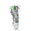
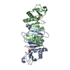
| Title | HIV-1 Nef protein in complex with single domain antibody sdAb19 and an engineered Hck SH3 domain | ||||||
 Components Components |
| ||||||
 Keywords Keywords | TRANSFERASE/APOPTOSIS/IMMUNE SYSTEM / SH3 domain / immunglobolin fold / Antibodies / Epitopes / HIV Antibodies / HIV Accessory Proteins / PxxP motif / Complementarity Determining Regions / TRANSFERASE-APOPTOSIS-IMMUNE SYSTEM complex | ||||||
| Function / homology |  Function and homology information Function and homology informationleukocyte degranulation / activation of transmembrane receptor protein tyrosine kinase activity / leukocyte migration involved in immune response / respiratory burst after phagocytosis / innate immune response-activating signaling pathway / symbiont-mediated suppression of host antigen processing and presentation of peptide antigen via MHC class I / symbiont-mediated suppression of host antigen processing and presentation of peptide antigen via MHC class II / regulation of podosome assembly / symbiont-mediated suppression of host autophagy / FLT3 signaling through SRC family kinases ...leukocyte degranulation / activation of transmembrane receptor protein tyrosine kinase activity / leukocyte migration involved in immune response / respiratory burst after phagocytosis / innate immune response-activating signaling pathway / symbiont-mediated suppression of host antigen processing and presentation of peptide antigen via MHC class I / symbiont-mediated suppression of host antigen processing and presentation of peptide antigen via MHC class II / regulation of podosome assembly / symbiont-mediated suppression of host autophagy / FLT3 signaling through SRC family kinases / regulation of phagocytosis / : / Nef and signal transduction / Fc-gamma receptor signaling pathway involved in phagocytosis / mesoderm development / positive regulation of actin filament polymerization / host cell Golgi membrane / FCGR activation / type II interferon-mediated signaling pathway / transport vesicle / Signaling by CSF3 (G-CSF) / phosphotyrosine residue binding / FCGR3A-mediated IL10 synthesis / peptidyl-tyrosine phosphorylation / cell surface receptor protein tyrosine kinase signaling pathway / lipopolysaccharide-mediated signaling pathway / cell projection / integrin-mediated signaling pathway / regulation of actin cytoskeleton organization / non-membrane spanning protein tyrosine kinase activity / FCGR3A-mediated phagocytosis / non-specific protein-tyrosine kinase / Regulation of signaling by CBL / negative regulation of inflammatory response to antigenic stimulus / caveola / Inactivation of CSF3 (G-CSF) signaling / SH3 domain binding / virion component / cytoplasmic side of plasma membrane / cytokine-mediated signaling pathway / Signaling by CSF1 (M-CSF) in myeloid cells / regulation of cell shape / protein autophosphorylation / regulation of inflammatory response / protein tyrosine kinase activity / cytoskeleton / cell differentiation / protein phosphorylation / lysosome / cell adhesion / intracellular signal transduction / defense response to Gram-positive bacterium / endocytosis involved in viral entry into host cell / inflammatory response / intracellular membrane-bounded organelle / signaling receptor binding / focal adhesion / positive regulation of cell population proliferation / lipid binding / negative regulation of apoptotic process / GTP binding / host cell plasma membrane / Golgi apparatus / extracellular region / ATP binding / membrane / nucleus / plasma membrane / cytosol Similarity search - Function | ||||||
| Biological species |  Homo sapiens (human) Homo sapiens (human) HIV-1 M:B_ARV2/SF2 (virus) HIV-1 M:B_ARV2/SF2 (virus) | ||||||
| Method |  X-RAY DIFFRACTION / X-RAY DIFFRACTION /  SYNCHROTRON / SYNCHROTRON /  MOLECULAR REPLACEMENT / Resolution: 2 Å MOLECULAR REPLACEMENT / Resolution: 2 Å | ||||||
 Authors Authors | Geyer, M. / Lulf, S. | ||||||
 Citation Citation |  Journal: Retrovirology / Year: 2014 Journal: Retrovirology / Year: 2014Title: Structural basis for the inhibition of HIV-1 Nef by a high-affinity binding single-domain antibody. Authors: Lulf, S. / Matz, J. / Rouyez, M.C. / Jarviluoma, A. / Saksela, K. / Benichou, S. / Geyer, M. | ||||||
| History |
|
- Structure visualization
| Structure viewer | Molecule:  Molmil Molmil Jmol/JSmol Jmol/JSmol |
|---|
- Downloads & links
- Download
| PDBx/mmCIF format |  4orz.cif.gz 4orz.cif.gz | 75.8 KB | Display |  PDBx/mmCIF format PDBx/mmCIF format |
|---|---|---|---|---|
| PDB format |  pdb4orz.ent.gz pdb4orz.ent.gz | 55.2 KB | Display |  PDB format PDB format |
| PDBx/mmJSON format |  4orz.json.gz 4orz.json.gz | Tree view |  PDBx/mmJSON format PDBx/mmJSON format | |
| Others |  Other downloads Other downloads |
-Validation report
| Arichive directory |  https://data.pdbj.org/pub/pdb/validation_reports/or/4orz https://data.pdbj.org/pub/pdb/validation_reports/or/4orz ftp://data.pdbj.org/pub/pdb/validation_reports/or/4orz ftp://data.pdbj.org/pub/pdb/validation_reports/or/4orz | HTTPS FTP |
|---|
-Related structure data
| Related structure data |  3rbbS S: Starting model for refinement |
|---|---|
| Similar structure data |
- Links
Links
- Assembly
| Deposited unit | 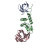
| ||||||||
|---|---|---|---|---|---|---|---|---|---|
| 1 |
| ||||||||
| Unit cell |
|
- Components
| #1: Protein | Mass: 7614.431 Da / Num. of mol.: 1 / Fragment: SH3 domain, UNP residues 77-138 / Mutation: E90Y, A91S, I92P, H93F, H94S, E95W Source method: isolated from a genetically manipulated source Source: (gene. exp.)  Homo sapiens (human) / Gene: HCK / Production host: Homo sapiens (human) / Gene: HCK / Production host:  References: UniProt: P08631, non-specific protein-tyrosine kinase |
|---|---|
| #2: Protein | Mass: 16817.979 Da / Num. of mol.: 1 / Fragment: Nef protein, UNP residues 45-210 / Mutation: I47M, T48A, C59S, C210A Source method: isolated from a genetically manipulated source Source: (gene. exp.)  HIV-1 M:B_ARV2/SF2 (virus) / Gene: nef / Production host: HIV-1 M:B_ARV2/SF2 (virus) / Gene: nef / Production host:  |
| #3: Antibody | Mass: 13007.562 Da / Num. of mol.: 1 Source method: isolated from a genetically manipulated source Source: (gene. exp.)   |
| #4: Water | ChemComp-HOH / |
-Experimental details
-Experiment
| Experiment | Method:  X-RAY DIFFRACTION / Number of used crystals: 1 X-RAY DIFFRACTION / Number of used crystals: 1 |
|---|
- Sample preparation
| Crystal | Density Matthews: 2.53 Å3/Da / Density % sol: 51.31 % |
|---|---|
| Crystal grow | Temperature: 293 K / Method: vapor diffusion, hanging drop / pH: 9 Details: About 0.1 micro L of protein solution at 10 mg/ml concentration was mixed with 0.1 micro L of reservoir solution from a 70 L reservoir in 96-well Hampton 3553 crystallization plates. Initial ...Details: About 0.1 micro L of protein solution at 10 mg/ml concentration was mixed with 0.1 micro L of reservoir solution from a 70 L reservoir in 96-well Hampton 3553 crystallization plates. Initial crystals of NefSF2 sdAb19 SH3B6 could be obtained in 0.2 M potassium formate and 20% polyethylene glycol (PEG) 3350. Crystal conditions were optimized to 0.2 M potassium formate, 17.5% polyethylene glycol (PEG) 3350 and 0.35 M ammonium chloride grown by hanging-drop vapor diffusion in Linbro crystallization plates. , pH 9.0, VAPOR DIFFUSION, HANGING DROP, temperature 293K |
-Data collection
| Diffraction | Mean temperature: 100 K |
|---|---|
| Diffraction source | Source:  SYNCHROTRON / Site: SYNCHROTRON / Site:  SLS SLS  / Beamline: X10SA / Wavelength: 0.9794 Å / Beamline: X10SA / Wavelength: 0.9794 Å |
| Detector | Type: PSI PILATUS 6M / Detector: PIXEL / Date: Feb 10, 2012 |
| Radiation | Monochromator: Diamond(111) / Protocol: SINGLE WAVELENGTH / Monochromatic (M) / Laue (L): M / Scattering type: x-ray |
| Radiation wavelength | Wavelength: 0.9794 Å / Relative weight: 1 |
| Reflection | Resolution: 2→41.75 Å / Num. obs: 47918 / % possible obs: 100 % / Observed criterion σ(F): -3 / Observed criterion σ(I): -3 |
| Reflection shell | Resolution: 2.1→2.15 Å / % possible all: 100 |
- Processing
| Software |
| ||||||||||||||||||||||||||||||||||||||||||||||||||||||||||||||||||||||
|---|---|---|---|---|---|---|---|---|---|---|---|---|---|---|---|---|---|---|---|---|---|---|---|---|---|---|---|---|---|---|---|---|---|---|---|---|---|---|---|---|---|---|---|---|---|---|---|---|---|---|---|---|---|---|---|---|---|---|---|---|---|---|---|---|---|---|---|---|---|---|---|
| Refinement | Method to determine structure:  MOLECULAR REPLACEMENT MOLECULAR REPLACEMENTStarting model: PDB ENTRY 3RBB Resolution: 2→41.75 Å / SU ML: 0.21 / σ(F): 2.04 / Phase error: 24.91 / Stereochemistry target values: MLHL
| ||||||||||||||||||||||||||||||||||||||||||||||||||||||||||||||||||||||
| Solvent computation | Shrinkage radii: 0.9 Å / VDW probe radii: 1.11 Å / Solvent model: FLAT BULK SOLVENT MODEL | ||||||||||||||||||||||||||||||||||||||||||||||||||||||||||||||||||||||
| Refinement step | Cycle: LAST / Resolution: 2→41.75 Å
| ||||||||||||||||||||||||||||||||||||||||||||||||||||||||||||||||||||||
| Refine LS restraints |
| ||||||||||||||||||||||||||||||||||||||||||||||||||||||||||||||||||||||
| LS refinement shell |
|
 Movie
Movie Controller







 PDBj
PDBj


