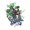
(3S)-3-amino-3-(3-chloro-4-hydroxyphenyl)propanoyl-[peptidyl-carrier protein SgcC2] monooxygenase / oxidoreductase activity, acting on the CH-CH group of donors / antibiotic biosynthetic process / monooxygenase activity Similarity search - Function
4-HPA 3-monooxygenase large component/Pyoverdin chromophore biosynthetic protein / HpaB/PvcC/4-BUDH / HpaB/PvcC/4-BUDH N-terminal / HpaB/PvcC/4-BUDH C-terminal / 4-hydroxyphenylacetate 3-hydroxylase C terminal / 4-hydroxyphenylacetate 3-hydroxylase N terminal / Butyryl-CoA Dehydrogenase, subunit A, domain 3 / Acyl-CoA oxidase/dehydrogenase, middle domain superfamily / Acyl-CoA dehydrogenase/oxidase, N-terminal and middle domain superfamily / Acyl-CoA dehydrogenase-like, C-terminal ...4-HPA 3-monooxygenase large component/Pyoverdin chromophore biosynthetic protein / HpaB/PvcC/4-BUDH / HpaB/PvcC/4-BUDH N-terminal / HpaB/PvcC/4-BUDH C-terminal / 4-hydroxyphenylacetate 3-hydroxylase C terminal / 4-hydroxyphenylacetate 3-hydroxylase N terminal / Butyryl-CoA Dehydrogenase, subunit A, domain 3 / Acyl-CoA oxidase/dehydrogenase, middle domain superfamily / Acyl-CoA dehydrogenase/oxidase, N-terminal and middle domain superfamily / Acyl-CoA dehydrogenase-like, C-terminal / Butyryl-CoA Dehydrogenase, subunit A; domain 3 / Up-down Bundle / Mainly Alpha Similarity search - Domain/homology
(3S)-3-amino-3-(3-chloro-4-hydroxyphenyl)propanoyl-[peptidyl-carrier protein SgcC2] monooxygenase Similarity search - Component
Mass: 18.015 Da / Num. of mol.: 217 / Source method: isolated from a natural source / Formula: H2O
Has protein modification
Y
-
Experimental details
-
Experiment
Experiment
Method: X-RAY DIFFRACTION / Number of used crystals: 1
-
Sample preparation
Crystal
Density Matthews: 2.08 Å3/Da / Density % sol: 40.91 %
Crystal grow
Temperature: 293.15 K / Method: vapor diffusion, hanging drop / pH: 6.5 Details: 1:1 v/v mixture of SgcC (15mg/mL) with reservoir solution (Molecular Dimensions Morpheus screen A1 condition: 60mM mixture of MgCl2 and CaCl2; 100mM Imidazole and MES buffer pH 6.5; 30% ...Details: 1:1 v/v mixture of SgcC (15mg/mL) with reservoir solution (Molecular Dimensions Morpheus screen A1 condition: 60mM mixture of MgCl2 and CaCl2; 100mM Imidazole and MES buffer pH 6.5; 30% mixture of PEG550mme and PEG 20k), cryoprotected by Mitegen LV cryo-oil, VAPOR DIFFUSION, HANGING DROP, temperature 293.15K
In the structure databanks used in Yorodumi, some data are registered as the other names, "COVID-19 virus" and "2019-nCoV". Here are the details of the virus and the list of structure data.
Jan 31, 2019. EMDB accession codes are about to change! (news from PDBe EMDB page)
EMDB accession codes are about to change! (news from PDBe EMDB page)
The allocation of 4 digits for EMDB accession codes will soon come to an end. Whilst these codes will remain in use, new EMDB accession codes will include an additional digit and will expand incrementally as the available range of codes is exhausted. The current 4-digit format prefixed with “EMD-” (i.e. EMD-XXXX) will advance to a 5-digit format (i.e. EMD-XXXXX), and so on. It is currently estimated that the 4-digit codes will be depleted around Spring 2019, at which point the 5-digit format will come into force.
The EM Navigator/Yorodumi systems omit the EMD- prefix.
Related info.:Q: What is EMD? / ID/Accession-code notation in Yorodumi/EM Navigator
Yorodumi is a browser for structure data from EMDB, PDB, SASBDB, etc.
This page is also the successor to EM Navigator detail page, and also detail information page/front-end page for Omokage search.
The word "yorodu" (or yorozu) is an old Japanese word meaning "ten thousand". "mi" (miru) is to see.
Related info.:EMDB / PDB / SASBDB / Comparison of 3 databanks / Yorodumi Search / Aug 31, 2016. New EM Navigator & Yorodumi / Yorodumi Papers / Jmol/JSmol / Function and homology information / Changes in new EM Navigator and Yorodumi
 Movie
Movie Controller


 Open data
 Basic information
 Components
 Keywords
 Function and homology information
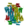
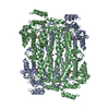
 Streptomyces globisporus (bacteria)
 X-RAY DIFFRACTION /
  Authors
 Citation
 Structure visualization
 Downloads & links
 Other downloads

 PDBj
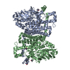
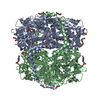
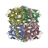
PDBj Assembly
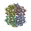

 Mass: 40.078 Da / Num. of mol.: 2 / Source method: obtained synthetically / Formula: Ca
 Mass: 92.094 Da / Num. of mol.: 1 / Source method: obtained synthetically / Formula: C3H8O3
 Mass: 18.015 Da / Num. of mol.: 217 / Source method: isolated from a natural source / Formula: H2O
 Sample preparation
 / Beamline: 23-ID-B / Wavelength: 0.91165 Å
 Processing