[English] 日本語
Yorodumi
- PDB-4obc: Crystal structure of HCV polymerase NS5b genotype 2a JFH-1 isolat... -

+
Open data


ID or keywords:

Loading...

-
Basic information

Entry
Database: PDB / ID: 4obc
Title
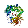
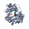
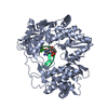
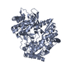
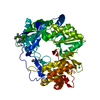
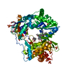
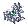
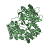
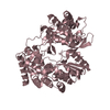
Crystal structure of HCV polymerase NS5b genotype 2a JFH-1 isolate with the S15G, C223H, V321I resistance mutations against the guanosine analog GS-0938 (PSI-3529238)
ComponentsRNA-directed RNA polymeraseRNA-dependent RNA polymerase
KeywordsViral Protein / transferase / Hepatitis / HCV / viral polymerase / RNA-dependent-RNA-polymerase / RdRp / resistance / nucleotide analog inhibitor
Function / homology
Function and homology information


negative regulation of autophagy of mitochondrion / : / hepacivirin / host cell mitochondrial membrane / host cell lipid droplet / symbiont-mediated suppression of host TRAF-mediated signal transduction / transformation of host cell by virus / Dectin-2 family / symbiont-mediated perturbation of host cell cycle G1/S transition checkpoint / symbiont-mediated suppression of host JAK-STAT cascade via inhibition of STAT1 activity ...negative regulation of autophagy of mitochondrion / : / hepacivirin / host cell mitochondrial membrane / host cell lipid droplet / symbiont-mediated suppression of host TRAF-mediated signal transduction / transformation of host cell by virus / Dectin-2 family / symbiont-mediated perturbation of host cell cycle G1/S transition checkpoint / symbiont-mediated suppression of host JAK-STAT cascade via inhibition of STAT1 activity / symbiont-mediated suppression of host cytoplasmic pattern recognition receptor signaling pathway via inhibition of MAVS activity / negative regulation of autophagy / SH3 domain binding / : / nucleoside-triphosphate phosphatase / protein complex oligomerization / monoatomic ion channel activity / clathrin-dependent endocytosis of virus by host cell / viral nucleocapsid / Hydrolases; Acting on peptide bonds (peptidases); Cysteine endopeptidases / RNA helicase activity / host cell endoplasmic reticulum membrane / host cell perinuclear region of cytoplasm / RNA helicase / induction by virus of host autophagy / ribonucleoprotein complex / RNA-directed RNA polymerase / viral RNA genome replication / cysteine-type endopeptidase activity / RNA-dependent RNA polymerase activity / serine-type endopeptidase activity / fusion of virus membrane with host endosome membrane / viral envelope / symbiont-mediated suppression of host type I interferon-mediated signaling pathway / host cell nucleus / structural molecule activity / virion attachment to host cell / host cell plasma membrane / virion membrane / ATP hydrolysis activity / proteolysis / RNA binding / zinc ion binding / ATP binding / membrane / identical protein binding
Similarity search - Function
Hepatitis C virus core protein, chain A superfamily / Hepatitus C virus, Non-structural 5a protein, C-terminal / Hepatitis C virus NS5A, 1B domain superfamily / Hepatitis C virus non-structural protein NS2, C-terminal domain / Hepatitis C virus non-structural protein NS2, N-terminal domain / Hepatitis C virus non-structural protein NS2 / HCV NS5a protein C-terminal region / Hepatitis C virus, Non-structural protein NS4b / Hepatitis C virus, Core protein, N-terminal / Hepatitis C virus non-structural protein NS4b ...Hepatitis C virus core protein, chain A superfamily / Hepatitus C virus, Non-structural 5a protein, C-terminal / Hepatitis C virus NS5A, 1B domain superfamily / Hepatitis C virus non-structural protein NS2, C-terminal domain / Hepatitis C virus non-structural protein NS2, N-terminal domain / Hepatitis C virus non-structural protein NS2 / HCV NS5a protein C-terminal region / Hepatitis C virus, Non-structural protein NS4b / Hepatitis C virus, Core protein, N-terminal / Hepatitis C virus non-structural protein NS4b / Hepatitis C virus capsid protein / Hepatitis C virus, Non-structural protein NS2 / Hepatitis C virus, Non-structural 5a protein / Hepatitis C virus, Non-structural 5a protein, domain 1a / Hepatitis C virus non-structural 5a, 1B domain / NS5A domain 1a superfamily / Hepatitis C virus non-structural 5a protein membrane anchor / Hepatitis C virus non-structural 5a zinc finger domain / Hepatitis C virus non-structural 5a domain 1b / Hepacivirus nonstructural protein 2 (NS2) protease domain profile. / Hepatitis C virus, Non-structural protein NS4a / Hepatitis C virus non-structural protein NS4a / Hepatitis C virus, Core protein, C-terminal / Hepatitis C virus core protein / Hepatitis C virus, Non-structural protein E2/NS1 / Hepatitis C virus non-structural protein E2/NS1 / Hepatitis C virus, Envelope glycoprotein E1 / Hepatitis C virus envelope glycoprotein E1 / RNA dependent RNA polymerase, hepatitis C virus / Viral RNA dependent RNA polymerase / Hepatitis C virus, NS3 protease, Peptidase S29 / Hepatitis C virus NS3 protease / Hepacivirus/Pegivirus NS3 protease domain profile. / DEAD box, Flavivirus / Flavivirus DEAD domain / Superfamilies 1 and 2 helicase C-terminal domain profile. / Superfamilies 1 and 2 helicase ATP-binding type-1 domain profile. / DEAD-like helicases superfamily / Helicase, C-terminal / Helicase superfamily 1/2, ATP-binding domain / Reverse transcriptase/Diguanylate cyclase domain / RNA-directed RNA polymerase, catalytic domain / RdRp of positive ssRNA viruses catalytic domain profile. / Peptidase S1, PA clan, chymotrypsin-like fold / Peptidase S1, PA clan / DNA/RNA polymerase superfamily / P-loop containing nucleoside triphosphate hydrolase
Similarity search - Domain/homology
Chem-PG6 / Genome polyprotein
Similarity search - Component
Biological speciesHepatitis C virus JFH-1
MethodX-RAY DIFFRACTION / SYNCHROTRON / MOLECULAR REPLACEMENT / molecular replacement / Resolution: 2.5 Å
AuthorsEdwards, T.E. / Abendroth, J. / Appleby, T.C.
CitationJournal: Antimicrob.Agents Chemother. / Year: 2014
Title: Molecular and Structural Basis for the Roles of Hepatitis C Virus Polymerase NS5B Amino Acids 15, 223, and 321 in Viral Replication and Drug Resistance.
Authors: Lam, A.M. / Edwards, T.E. / Mosley, R.T. / Murakami, E. / Bansal, S. / Lugo, C. / Bao, H. / Otto, M.J. / Sofia, M.J. / Furman, P.A.
History
DepositionJan 7, 2014Deposition site: RCSB / Processing site: RCSB
Revision 1.0Sep 10, 2014Provider: repository / Type: Initial release
Revision 1.1Oct 29, 2014Group: Database references
Revision 1.2Nov 22, 2017Group: Refinement description / Category: software
Revision 1.3Sep 20, 2023Group: Data collection / Database references ...Data collection / Database references / Derived calculations / Refinement description
Category: chem_comp_atom / chem_comp_bond ...chem_comp_atom / chem_comp_bond / database_2 / pdbx_initial_refinement_model / struct_ref_seq_dif / struct_site
Item: _database_2.pdbx_DOI / _database_2.pdbx_database_accession ..._database_2.pdbx_DOI / _database_2.pdbx_database_accession / _struct_ref_seq_dif.details / _struct_site.pdbx_auth_asym_id / _struct_site.pdbx_auth_comp_id / _struct_site.pdbx_auth_seq_id

-
Structure visualization

Structure viewerMolecule:
MolmilJmol/JSmol

Downloads & links

-
Assembly

Deposited unit
A: RNA-directed RNA polymerase
hetero molecules


Theoretical massNumber of molelcules
Total (without water)65,5016
Polymers64,8191
Non-polymers6815
Water3,711206
1


  • Idetical with deposited unit
  • defined by author&software
TypeNameSymmetry operationNumber
identity operation1_555x,y,z1
MethodPISA
Unit cell
Length a, b, c (Å)140.208, 140.208, 92.567
Angle α, β, γ (deg.)90.000, 90.000, 120.000
Int Tables number170
Space group name H-MP65

-
Components

-
Protein , 1 types, 1 molecules A

#1: Protein RNA-directed RNA polymerase / RNA-dependent RNA polymerase / NS5B / p68


Mass: 64819.379 Da / Num. of mol.: 1 / Mutation: S15G E86Q E87Q C223H V321I
Source method: isolated from a genetically manipulated source
Source: (gene. exp.) Hepatitis C virus JFH-1 / Strain: JFH-1 / Gene: POLG_HCVJF / Plasmid: V5854 / Production host: Escherichia coli (E. coli) / Strain (production host): Rosetta DE3 / References: UniProt: Q99IB8, RNA-directed RNA polymerase

-
Non-polymers , 5 types, 211 molecules

#2: Chemical ChemComp-MES / 2-(N-MORPHOLINO)-ETHANESULFONIC ACID / MES (buffer)


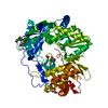
Mass: 195.237 Da / Num. of mol.: 1 / Source method: obtained synthetically / Formula: C6H13NO4S / Comment: pH buffer*YM
#3: Chemical ChemComp-GOL / GLYCEROL / GLYCERIN / PROPANE-1,2,3-TRIOL / Glycerol


Mass: 92.094 Da / Num. of mol.: 2 / Source method: obtained synthetically / Formula: C3H8O3
#4: Chemical ChemComp-PG6 / 1-(2-METHOXY-ETHOXY)-2-{2-[2-(2-METHOXY-ETHOXY]-ETHOXY}-ETHANE / Polyethylene glycol


Mass: 266.331 Da / Num. of mol.: 1 / Source method: obtained synthetically / Formula: C12H26O6
#5: Chemical ChemComp-CL / CHLORIDE ION / Chloride


Mass: 35.453 Da / Num. of mol.: 1 / Source method: obtained synthetically / Formula: Cl
#6: Water ChemComp-HOH / water / Water


Mass: 18.015 Da / Num. of mol.: 206 / Source method: isolated from a natural source / Formula: H2O

-
Experimental details

-
Experiment

ExperimentMethod: X-RAY DIFFRACTION / Number of used crystals: 1

-
Sample preparation

CrystalDensity Matthews: 4.05 Å3/Da / Density % sol: 69.64 %
Crystal growTemperature: 289 K / Method: vapor diffusion, sitting drop / pH: 8
Details: NS5b VCID 5854 at 4.25 mg/mL in 5 mM Tris pH 7.5, 200 mM NH4OAc, 1 mM EDTA 1 mM DTT against PACT screen condition D5, 25% PEG 1500, 0.1M MMT Malic Acid, MES, Tris Buffer pH 8.0 supplemented ...Details: NS5b VCID 5854 at 4.25 mg/mL in 5 mM Tris pH 7.5, 200 mM NH4OAc, 1 mM EDTA 1 mM DTT against PACT screen condition D5, 25% PEG 1500, 0.1M MMT Malic Acid, MES, Tris Buffer pH 8.0 supplemented with 20% glycerol as cryo-protectant, crystal tracking ID 223572d5, puck ID cps0237-1, VAPOR DIFFUSION, SITTING DROP, temperature 289K

-
Data collection

DiffractionMean temperature: 100 K
Diffraction sourceSource: SYNCHROTRON / Site: SSRL / Beamline: BL9-2 / Wavelength: 0.97946 Å
DetectorType: MARMOSAIC 325 mm CCD / Detector: CCD / Date: Jun 30, 2011
RadiationProtocol: SINGLE WAVELENGTH / Monochromatic (M) / Laue (L): M / Scattering type: x-ray
Radiation wavelengthWavelength: 0.97946 Å / Relative weight: 1
ReflectionResolution: 2.5→50 Å / Num. obs: 35841 / % possible obs: 99.7 % / Redundancy: 7.8 % / Rmerge(I) obs: 0.085 / Χ2: 0.99 / Net I/σ(I): 21.9
Reflection shell

Diffraction-ID: 1

Resolution (Å)Redundancy (%)Rmerge(I) obsMean I/σ(I) obsNum. unique allΧ2% possible all
2.5-2.573.70.6382.328770.95597.3
2.57-2.664.50.52129710.95799.3
2.66-2.755.60.43129930.991100
2.75-2.867.50.32429580.966100
2.86-2.9990.25130120.986100
2.99-3.1590.18129561.009100
3.15-3.3590.12829870.962100
3.35-3.6190.08829950.958100
3.61-3.9790.06929810.977100
3.97-4.5490.05530301.045100
4.54-5.7290.05330001.026100
5.72-508.80.04630811.00399.7

-
Phasing

PhasingMethod: molecular replacement
Phasing MRRfactor: 50.11 / Model details: Phaser MODE: MR_AUTO
Highest resolutionLowest resolution
Rotation3 Å41.12 Å
Translation3 Å41.12 Å

-
Processing

Software
NameVersionClassificationNB
DENZOdata reduction
SCALEPACKdata scaling
PHASER2.1.4phasing
REFMAC5.5.0109refinement
PDB_EXTRACT3.14data extraction
HKL-2000data reduction
HKL-2000data scaling
RefinementMethod to determine structure: MOLECULAR REPLACEMENT
Starting model: PDB ENTRY 1YUY
Resolution: 2.5→50 Å / Cor.coef. Fo:Fc: 0.948 / Cor.coef. Fo:Fc free: 0.931 / WRfactor Rfree: 0.209 / WRfactor Rwork: 0.1769 / Occupancy max: 1 / Occupancy min: 0.5 / FOM work R set: 0.8503 / SU B: 6.803 / SU ML: 0.151 / SU R Cruickshank DPI: 0.2565 / SU Rfree: 0.2079 / Cross valid method: THROUGHOUT / σ(F): 0 / ESU R: 0.257 / ESU R Free: 0.208 / Stereochemistry target values: MAXIMUM LIKELIHOOD
Details: HYDROGENS HAVE BEEN ADDED IN THE RIDING POSITIONS U VALUES : REFINED INDIVIDUALLY
RfactorNum. reflection% reflectionSelection details
Rfree0.2234 1792 5 %RANDOM
Rwork0.1882 ---
obs0.1899 35745 99.58 %-
Solvent computationIon probe radii: 0.8 Å / Shrinkage radii: 0.8 Å / VDW probe radii: 1.4 Å / Solvent model: MASK
Displacement parametersBiso max: 86.42 Å2 / Biso mean: 39.3253 Å2 / Biso min: 18.41 Å2
Baniso -1Baniso -2Baniso -3
1-1.88 Å20.94 Å20 Å2
2--1.88 Å20 Å2
3----2.81 Å2
Refinement stepCycle: LAST / Resolution: 2.5→50 Å
ProteinNucleic acidLigandSolventTotal
Num. atoms4277 0 43 206 4526
Refine LS restraints
Refine-IDTypeDev idealDev ideal targetNumber
X-RAY DIFFRACTIONr_bond_refined_d0.0130.0224426
X-RAY DIFFRACTIONr_angle_refined_deg1.4211.9726017
X-RAY DIFFRACTIONr_dihedral_angle_1_deg5.8485555
X-RAY DIFFRACTIONr_dihedral_angle_2_deg33.8522.216176
X-RAY DIFFRACTIONr_dihedral_angle_3_deg15.99815713
X-RAY DIFFRACTIONr_dihedral_angle_4_deg14.8591539
X-RAY DIFFRACTIONr_chiral_restr0.0870.2682
X-RAY DIFFRACTIONr_gen_planes_refined0.0060.0213311
X-RAY DIFFRACTIONr_mcbond_it0.7411.52776
X-RAY DIFFRACTIONr_mcangle_it1.45224482
X-RAY DIFFRACTIONr_scbond_it2.131650
X-RAY DIFFRACTIONr_scangle_it3.5854.51534
LS refinement shellResolution: 2.5→2.565 Å / Total num. of bins used: 20
RfactorNum. reflection% reflection
Rfree0.335 140 -
Rwork0.263 2400 -
all-2540 -
obs--97.32 %

+
About Yorodumi

-
News

-
Feb 9, 2022. New format data for meta-information of EMDB entries

New format data for meta-information of EMDB entries

  • Version 3 of the EMDB header file is now the official format.
  • The previous official version 1.9 will be removed from the archive.

Related info.:EMDB header

External links:wwPDB to switch to version 3 of the EMDB data model

-
Aug 12, 2020. Covid-19 info

Covid-19 info

URL: https://pdbj.org/emnavi/covid19.php

New page: Covid-19 featured information page in EM Navigator.

Related info.:Covid-19 info / Mar 5, 2020. Novel coronavirus structure data

+
Mar 5, 2020. Novel coronavirus structure data

Novel coronavirus structure data

Related info.:Yorodumi Speices / Aug 12, 2020. Covid-19 info

External links:COVID-19 featured content - PDBj / Molecule of the Month (242):Coronavirus Proteases

+
Jan 31, 2019. EMDB accession codes are about to change! (news from PDBe EMDB page)

EMDB accession codes are about to change! (news from PDBe EMDB page)

  • The allocation of 4 digits for EMDB accession codes will soon come to an end. Whilst these codes will remain in use, new EMDB accession codes will include an additional digit and will expand incrementally as the available range of codes is exhausted. The current 4-digit format prefixed with “EMD-” (i.e. EMD-XXXX) will advance to a 5-digit format (i.e. EMD-XXXXX), and so on. It is currently estimated that the 4-digit codes will be depleted around Spring 2019, at which point the 5-digit format will come into force.
  • The EM Navigator/Yorodumi systems omit the EMD- prefix.

Related info.:Q: What is EMD? / ID/Accession-code notation in Yorodumi/EM Navigator

External links:EMDB Accession Codes are Changing Soon! / Contact to PDBj

+
Jul 12, 2017. Major update of PDB

Major update of PDB

  • wwPDB released updated PDB data conforming to the new PDBx/mmCIF dictionary.
  • This is a major update changing the version number from 4 to 5, and with Remediation, in which all the entries are updated.
  • In this update, many items about electron microscopy experimental information are reorganized (e.g. em_software).
  • Now, EM Navigator and Yorodumi are based on the updated data.

External links:wwPDB Remediation / Enriched Model Files Conforming to OneDep Data Standards Now Available in the PDB FTP Archive

-
Yorodumi

Thousand views of thousand structures

  • Yorodumi is a browser for structure data from EMDB, PDB, SASBDB, etc.
  • This page is also the successor to EM Navigator detail page, and also detail information page/front-end page for Omokage search.
  • The word "yorodu" (or yorozu) is an old Japanese word meaning "ten thousand". "mi" (miru) is to see.

Related info.:EMDB / PDB / SASBDB / Comparison of 3 databanks / Yorodumi Search / Aug 31, 2016. New EM Navigator & Yorodumi / Yorodumi Papers / Jmol/JSmol / Function and homology information / Changes in new EM Navigator and Yorodumi

Read more