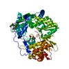
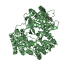
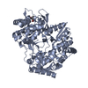
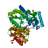
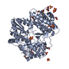
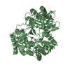
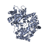
[English] 日本語
Yorodumi
- PDB-5qj1: CRYSTAL STRUCTURE OF THE HEPATITIS C VIRUS GENOTYPE 2A STRAIN JFH... -

+
Open data


ID or keywords:

Loading...

-
Basic information

Entry
Database: PDB / ID: 5qj1
TitleCRYSTAL STRUCTURE OF THE HEPATITIS C VIRUS GENOTYPE 2A STRAIN JFH1 L30S NS5B RNA-DEPENDENT RNA POLYMERASE IN COMPLEX WITH 6-(ethylamino)-2-(4-fluorophenyl)-5-(3-{[1-(5-fluoropyrimidin-2-yl)cyclopropyl]carbamoyl}-4-methoxyphenyl)-N-methyl-1-benzofuran-3-carboxamide
ComponentsRNA-dependent RNA polymerase
KeywordsTRANSFERASE/TRANSFERASE inhibitor / RDRP STRUCTURE (FINGERS / PALM / THUMB DOMAINS) / ACETYLATION / APOPTOSIS / ATP-BINDING / CAPSID PROTEIN / CELL MEMBRANE / CYTOPLASM / DISULFIDE BOND / ENDOPLASMIC RETICULUM / ENVELOPE PROTEIN / FUSION PROTEIN / GLYCOPROTEIN / HELICASE / HOST-VIRUS INTERACTION / HYDROLASE / INTERFERON ANTIVIRAL SYSTEM EVASION / LIPID DROPLET / LIPOPROTEIN / MEMBRANE / METAL- BINDING / MITOCHONDRION / MULTIFUNCTIONAL ENZYME / NUCLEOTIDE- BINDING / NUCLEOTIDYLTRANSFERASE / NUCLEUS / ONCOGENE / PALMITATE / PHOSPHOPROTEIN / PROTEASE / RIBONUCLEOPROTEIN / RNA REPLICATION / RNA-BINDING / RNA-DIRECTED RNA POLYMERASE / SECRETED / SERINE PROTEASE / SH3-BINDING / THIOL PROTEASE / TRANSCRIPTION / TRANSCRIPTION REGULATION / TRANSFERASE / TRANSMEMBRANE / UBL CONJUGATION / VIRAL NUCLEOPROTEIN / VIRION / ZINC / TRANSFERASE-TRANSFERASE inhibitor complex
Function / homology
Function and homology information


negative regulation of autophagy of mitochondrion / : / hepacivirin / host cell mitochondrial membrane / host cell lipid droplet / symbiont-mediated suppression of host TRAF-mediated signal transduction / transformation of host cell by virus / Dectin-2 family / symbiont-mediated perturbation of host cell cycle G1/S transition checkpoint / symbiont-mediated suppression of host JAK-STAT cascade via inhibition of STAT1 activity ...negative regulation of autophagy of mitochondrion / : / hepacivirin / host cell mitochondrial membrane / host cell lipid droplet / symbiont-mediated suppression of host TRAF-mediated signal transduction / transformation of host cell by virus / Dectin-2 family / symbiont-mediated perturbation of host cell cycle G1/S transition checkpoint / symbiont-mediated suppression of host JAK-STAT cascade via inhibition of STAT1 activity / symbiont-mediated suppression of host cytoplasmic pattern recognition receptor signaling pathway via inhibition of MAVS activity / ribonucleoside triphosphate phosphatase activity / lipid droplet / negative regulation of autophagy / SH3 domain binding / : / nucleoside-triphosphate phosphatase / protein complex oligomerization / monoatomic ion channel activity / clathrin-dependent endocytosis of virus by host cell / viral nucleocapsid / Hydrolases; Acting on peptide bonds (peptidases); Cysteine endopeptidases / RNA helicase activity / host cell endoplasmic reticulum membrane / host cell perinuclear region of cytoplasm / RNA helicase / induction by virus of host autophagy / ribonucleoprotein complex / RNA-directed RNA polymerase / viral RNA genome replication / cysteine-type endopeptidase activity / RNA-dependent RNA polymerase activity / serine-type endopeptidase activity / fusion of virus membrane with host endosome membrane / viral envelope / symbiont-mediated suppression of host type I interferon-mediated signaling pathway / host cell nucleus / virion attachment to host cell / host cell plasma membrane / virion membrane / structural molecule activity / ATP hydrolysis activity / proteolysis / RNA binding / zinc ion binding / ATP binding / membrane / identical protein binding / plasma membrane / cytoplasm
Similarity search - Function
Hepatitis C virus core protein, chain A superfamily / Hepatitus C virus, Non-structural 5a protein, C-terminal / Hepatitis C virus NS5A, 1B domain superfamily / Hepatitis C virus non-structural protein NS2, C-terminal domain / Hepatitis C virus non-structural protein NS2, N-terminal domain / Hepatitis C virus non-structural protein NS2 / HCV NS5a protein C-terminal region / Hepatitis C virus, Non-structural protein NS4b / Hepatitis C virus, Core protein, N-terminal / Hepatitis C virus non-structural protein NS4b ...Hepatitis C virus core protein, chain A superfamily / Hepatitus C virus, Non-structural 5a protein, C-terminal / Hepatitis C virus NS5A, 1B domain superfamily / Hepatitis C virus non-structural protein NS2, C-terminal domain / Hepatitis C virus non-structural protein NS2, N-terminal domain / Hepatitis C virus non-structural protein NS2 / HCV NS5a protein C-terminal region / Hepatitis C virus, Non-structural protein NS4b / Hepatitis C virus, Core protein, N-terminal / Hepatitis C virus non-structural protein NS4b / Hepatitis C virus capsid protein / Hepatitis C virus, Non-structural protein NS2 / Hepatitis C virus, Non-structural 5a protein / Hepatitis C virus, Non-structural 5a protein, domain 1a / Hepatitis C virus non-structural 5a, 1B domain / NS5A domain 1a superfamily / Hepatitis C virus non-structural 5a protein membrane anchor / Hepatitis C virus non-structural 5a zinc finger domain / Hepatitis C virus non-structural 5a domain 1b / Hepacivirus nonstructural protein 2 (NS2) protease domain profile. / Hepatitis C virus, Non-structural protein NS4a / Hepatitis C virus non-structural protein NS4a / Hepatitis C virus, Core protein, C-terminal / Hepatitis C virus core protein / Hepatitis C virus, Non-structural protein E2/NS1 / Hepatitis C virus non-structural protein E2/NS1 / Hepatitis C virus, Envelope glycoprotein E1 / Hepatitis C virus envelope glycoprotein E1 / RNA dependent RNA polymerase, hepatitis C virus / Viral RNA dependent RNA polymerase / Hepatitis C virus, NS3 protease, Peptidase S29 / Hepatitis C virus NS3 protease / Hepacivirus/Pegivirus NS3 protease domain profile. / DEAD box, Flavivirus / Flavivirus DEAD domain / Superfamilies 1 and 2 helicase C-terminal domain profile. / Superfamilies 1 and 2 helicase ATP-binding type-1 domain profile. / DEAD-like helicases superfamily / Helicase, C-terminal / Helicase superfamily 1/2, ATP-binding domain / Reverse transcriptase/Diguanylate cyclase domain / RNA-directed RNA polymerase, catalytic domain / RdRp of positive ssRNA viruses catalytic domain profile. / Peptidase S1, PA clan, chymotrypsin-like fold / Peptidase S1, PA clan / DNA/RNA polymerase superfamily / P-loop containing nucleoside triphosphate hydrolase
Similarity search - Domain/homology
Chem-J6J / Genome polyprotein / Genome polyprotein
Similarity search - Component
Biological speciesHepacivirus C
MethodX-RAY DIFFRACTION / SYNCHROTRON / MOLECULAR REPLACEMENT / molecular replacement / Resolution: 2.17 Å
AuthorsSheriff, S.
CitationJournal: ACS Med Chem Lett / Year: 2018
Title: Structure-Property Basis for Solving Transporter-Mediated Efflux and Pan-Genotypic Inhibition in HCV NS5B Inhibitors.
Authors: Yeung, K.S. / Beno, B.R. / Mosure, K. / Zhu, J. / Grant-Young, K.A. / Parcella, K. / Anjanappa, P. / Bora, R.O. / Selvakumar, K. / Wang, Y.K. / Fang, H. / Krause, R. / Rigat, K. / Liu, M. / ...Authors: Yeung, K.S. / Beno, B.R. / Mosure, K. / Zhu, J. / Grant-Young, K.A. / Parcella, K. / Anjanappa, P. / Bora, R.O. / Selvakumar, K. / Wang, Y.K. / Fang, H. / Krause, R. / Rigat, K. / Liu, M. / Lemm, J. / Sheriff, S. / Witmer, M. / Tredup, J. / Jardel, A. / Kish, K. / Parker, D. / Haskell, R. / Santone, K. / Meanwell, N.A. / Soars, M.G. / Roberts, S.B. / Kadow, J.F.
History
DepositionAug 13, 2018Deposition site: RCSB / Processing site: RCSB
Revision 1.0Nov 21, 2018Provider: repository / Type: Initial release
Revision 1.1Jan 16, 2019Group: Data collection / Database references / Category: citation / citation_author
Item: _citation.journal_abbrev / _citation.journal_volume ..._citation.journal_abbrev / _citation.journal_volume / _citation.page_first / _citation.page_last / _citation.pdbx_database_id_PubMed / _citation.title
Revision 1.2May 12, 2021Group: Structure summary / Category: pdbx_deposit_group / Item: _pdbx_deposit_group.group_type
Revision 1.3Mar 6, 2024Group: Data collection / Database references / Category: chem_comp_atom / chem_comp_bond / database_2
Item: _database_2.pdbx_DOI / _database_2.pdbx_database_accession

-
Structure visualization

Structure viewerMolecule:
MolmilJmol/JSmol

Downloads & links

-
Assembly

Deposited unit
A: RNA-dependent RNA polymerase
hetero molecules


Theoretical massNumber of molelcules
Total (without water)66,44519
Polymers63,9311
Non-polymers2,51318
Water5,801322
1


  • Idetical with deposited unit
  • defined by author
TypeNameSymmetry operationNumber
identity operation1_555x,y,z1
Unit cell
Length a, b, c (Å)93.440, 116.550, 64.710
Angle α, β, γ (deg.)90.000, 90.000, 90.000
Int Tables number18
Space group name H-MP21212
Components on special symmetry positions
IDModelComponents
11A-602-

SO4

-
Components

-
Protein , 1 types, 1 molecules A

#1: Protein RNA-dependent RNA polymerase /


Mass: 63931.406 Da / Num. of mol.: 1
Fragment: N-TERMINAL CATALYTIC REGION, UNP RESIDUES 2443- 3007
Mutation: L30S
Source method: isolated from a genetically manipulated source
Source: (gene. exp.) Hepacivirus C / Strain: 1 / Plasmid: PET21b / Production host: Escherichia coli BL21(DE3) (bacteria) / Strain (production host): BL21(DE3)
References: UniProt: R9THT8, UniProt: Q99IB8*PLUS, RNA-directed RNA polymerase

-
Non-polymers , 5 types, 340 molecules

#2: Chemical ChemComp-J6J / 6-(ethylamino)-2-(4-fluorophenyl)-5-(3-{[1-(5-fluoropyrimidin-2-yl)cyclopropyl]carbamoyl}-4-methoxyphenyl)-N-methyl-1-benzofuran-3-carboxamide


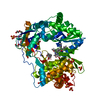
Mass: 597.611 Da / Num. of mol.: 1 / Source method: obtained synthetically / Formula: C33H29F2N5O4
#3: Chemical
ChemComp-SO4 / SULFATE ION / Sulfate


Mass: 96.063 Da / Num. of mol.: 11 / Source method: obtained synthetically / Formula: SO4
#4: Chemical ChemComp-GOL / GLYCEROL / GLYCERIN / PROPANE-1,2,3-TRIOL / Glycerol


Mass: 92.094 Da / Num. of mol.: 3 / Source method: obtained synthetically / Formula: C3H8O3
#5: Chemical ChemComp-PG4 / TETRAETHYLENE GLYCOL / Polyethylene glycol


Mass: 194.226 Da / Num. of mol.: 3 / Source method: obtained synthetically / Formula: C8H18O5 / Comment: precipitant*YM
#6: Water ChemComp-HOH / water / Water


Mass: 18.015 Da / Num. of mol.: 322 / Source method: isolated from a natural source / Formula: H2O

-
Experimental details

-
Experiment

ExperimentMethod: X-RAY DIFFRACTION / Number of used crystals: 1

-
Sample preparation

CrystalDensity Matthews: 2.76 Å3/Da / Density % sol: 55.37 %
Crystal growTemperature: 277 K / Method: vapor diffusion, hanging drop / pH: 4.6
Details: 100 mM sodium citrate, pH 4.6, 200 mM (NH4)2SO4, 25% (w/v) PEG 4000

-
Data collection

DiffractionMean temperature: 100 K
Diffraction sourceSource: SYNCHROTRON / Site: APS / Beamline: 17-ID / Wavelength: 1 / Wavelength: 1 Å
DetectorType: DECTRIS PILATUS 6M / Detector: PIXEL / Date: Apr 13, 2012
RadiationProtocol: SINGLE WAVELENGTH / Monochromatic (M) / Laue (L): M / Scattering type: x-ray
Radiation wavelength
IDWavelength (Å)Relative weight
111
211
ReflectionResolution: 2.17→46.72 Å / Num. obs: 38057 / % possible obs: 99.8 % / Observed criterion σ(I): 0 / Redundancy: 6.5 % / Biso Wilson estimate: 30.22 Å2 / Rsym value: 0.114 / Net I/σ(I): 13.1
Reflection shellResolution: 2.17→2.29 Å / Redundancy: 6.7 % / Mean I/σ(I) obs: 3.8 / Rsym value: 0.544 / Rejects: 0 / % possible all: 99.8

-
Phasing

PhasingMethod: molecular replacement

-
Processing

Software
NameVersionClassificationNB
XDSdata reduction
SCALAdata scaling
AMoREphasing
BUSTER2.11.7refinement
PDB_EXTRACT3.22data extraction
RefinementMethod to determine structure: MOLECULAR REPLACEMENT
Starting model: 1YUY
Resolution: 2.17→21.32 Å / Cor.coef. Fo:Fc: 0.938 / Cor.coef. Fo:Fc free: 0.915 / SU R Cruickshank DPI: 0.19 / Cross valid method: THROUGHOUT / σ(F): 0 / SU R Blow DPI: 0.201 / SU Rfree Blow DPI: 0.163 / SU Rfree Cruickshank DPI: 0.16
RfactorNum. reflection% reflectionSelection details
Rfree0.21 1898 5 %RANDOM
Rwork0.177 ---
obs0.179 37962 99.7 %-
Displacement parametersBiso max: 125.47 Å2 / Biso mean: 29.74 Å2 / Biso min: 10.55 Å2
Baniso -1Baniso -2Baniso -3
1-2.9187 Å20 Å20 Å2
2---4.593 Å20 Å2
3---1.6743 Å2
Refine analyzeLuzzati coordinate error obs: 0.24 Å
Refinement stepCycle: final / Resolution: 2.17→21.32 Å
ProteinNucleic acidLigandSolventTotal
Num. atoms4203 0 185 322 4710
Biso mean--41.85 39.31 -
Num. residues----543
Refine LS restraints
Refine-IDTypeNumberRestraint functionWeightDev ideal
X-RAY DIFFRACTIONt_dihedral_angle_d1522SINUSOIDAL2
X-RAY DIFFRACTIONt_trig_c_planes
X-RAY DIFFRACTIONt_gen_planes775HARMONIC5
X-RAY DIFFRACTIONt_it4478HARMONIC20
X-RAY DIFFRACTIONt_nbd
X-RAY DIFFRACTIONt_improper_torsion
X-RAY DIFFRACTIONt_pseud_angle
X-RAY DIFFRACTIONt_chiral_improper_torsion578SEMIHARMONIC5
X-RAY DIFFRACTIONt_sum_occupancies
X-RAY DIFFRACTIONt_utility_distance
X-RAY DIFFRACTIONt_utility_angle
X-RAY DIFFRACTIONt_utility_torsion
X-RAY DIFFRACTIONt_ideal_dist_contact5342SEMIHARMONIC4
X-RAY DIFFRACTIONt_bond_d4478HARMONIC20.01
X-RAY DIFFRACTIONt_angle_deg6102HARMONIC21.02
X-RAY DIFFRACTIONt_omega_torsion3.36
X-RAY DIFFRACTIONt_other_torsion16.98
LS refinement shellResolution: 2.17→2.23 Å / Total num. of bins used: 19
RfactorNum. reflection% reflection
Rfree0.2203 151 5.16 %
Rwork0.1875 2777 -
all0.1893 2928 -
obs--99.29 %

+
About Yorodumi

-
News

-
Feb 9, 2022. New format data for meta-information of EMDB entries

New format data for meta-information of EMDB entries

  • Version 3 of the EMDB header file is now the official format.
  • The previous official version 1.9 will be removed from the archive.

Related info.:EMDB header

External links:wwPDB to switch to version 3 of the EMDB data model

-
Aug 12, 2020. Covid-19 info

Covid-19 info

URL: https://pdbj.org/emnavi/covid19.php

New page: Covid-19 featured information page in EM Navigator.

Related info.:Covid-19 info / Mar 5, 2020. Novel coronavirus structure data

+
Mar 5, 2020. Novel coronavirus structure data

Novel coronavirus structure data

Related info.:Yorodumi Speices / Aug 12, 2020. Covid-19 info

External links:COVID-19 featured content - PDBj / Molecule of the Month (242):Coronavirus Proteases

+
Jan 31, 2019. EMDB accession codes are about to change! (news from PDBe EMDB page)

EMDB accession codes are about to change! (news from PDBe EMDB page)

  • The allocation of 4 digits for EMDB accession codes will soon come to an end. Whilst these codes will remain in use, new EMDB accession codes will include an additional digit and will expand incrementally as the available range of codes is exhausted. The current 4-digit format prefixed with “EMD-” (i.e. EMD-XXXX) will advance to a 5-digit format (i.e. EMD-XXXXX), and so on. It is currently estimated that the 4-digit codes will be depleted around Spring 2019, at which point the 5-digit format will come into force.
  • The EM Navigator/Yorodumi systems omit the EMD- prefix.

Related info.:Q: What is EMD? / ID/Accession-code notation in Yorodumi/EM Navigator

External links:EMDB Accession Codes are Changing Soon! / Contact to PDBj

+
Jul 12, 2017. Major update of PDB

Major update of PDB

  • wwPDB released updated PDB data conforming to the new PDBx/mmCIF dictionary.
  • This is a major update changing the version number from 4 to 5, and with Remediation, in which all the entries are updated.
  • In this update, many items about electron microscopy experimental information are reorganized (e.g. em_software).
  • Now, EM Navigator and Yorodumi are based on the updated data.

External links:wwPDB Remediation / Enriched Model Files Conforming to OneDep Data Standards Now Available in the PDB FTP Archive

-
Yorodumi

Thousand views of thousand structures

  • Yorodumi is a browser for structure data from EMDB, PDB, SASBDB, etc.
  • This page is also the successor to EM Navigator detail page, and also detail information page/front-end page for Omokage search.
  • The word "yorodu" (or yorozu) is an old Japanese word meaning "ten thousand". "mi" (miru) is to see.

Related info.:EMDB / PDB / SASBDB / Comparison of 3 databanks / Yorodumi Search / Aug 31, 2016. New EM Navigator & Yorodumi / Yorodumi Papers / Jmol/JSmol / Function and homology information / Changes in new EM Navigator and Yorodumi

Read more