Mass: 18.015 Da / Num. of mol.: 169 / Source method: isolated from a natural source / Formula: H2O
Sequence details
PROTEIN COMPRISES UNP RESIDUES 2443-3012 WITH RESIDUES 2886-2895 REPLACED WITH A GG LINKER.
-
Experimental details
-
Experiment
Experiment
Method: X-RAY DIFFRACTION / Number of used crystals: 1
-
Sample preparation
Crystal
Density Matthews: 4.15 Å3/Da / Density % sol: 70.38 %
Crystal grow
Temperature: 289 K / Method: vapor diffusion, sitting drop / pH: 6.9 Details: 3.95 mg/mL NS5B in 20 mM Tris, pH 8, 200 mM sodium chloride, 20% glycerol, 2 mM TCEP, 200 mM imidazole against 30% PEG550 MME, 0.1 M Bis-Tris propane, pH 6.5, 50 mM ammonium sulfate, ...Details: 3.95 mg/mL NS5B in 20 mM Tris, pH 8, 200 mM sodium chloride, 20% glycerol, 2 mM TCEP, 200 mM imidazole against 30% PEG550 MME, 0.1 M Bis-Tris propane, pH 6.5, 50 mM ammonium sulfate, cryoprotectant: 25% ethylene glycol, crystal tracking ID 227386E9, VAPOR DIFFUSION, SITTING DROP, temperature 289K
-
Data collection
Diffraction
Mean temperature: 100 K
Diffraction source
Source: SYNCHROTRON / Site: ALS / Beamline: 5.0.3 / Wavelength: 0.9765 / Wavelength: 0.9765 Å
Resolution: 2.5→50 Å / Cor.coef. Fo:Fc: 0.943 / Cor.coef. Fo:Fc free: 0.918 / WRfactor Rfree: 0.1863 / WRfactor Rwork: 0.1507 / Occupancy max: 1 / Occupancy min: 1 / FOM work R set: 0.7625 / SU B: 19.29 / SU ML: 0.191 / SU R Cruickshank DPI: 0.297 / SU Rfree: 0.2439 / Cross valid method: THROUGHOUT / σ(F): 0 / ESU R: 0.297 / ESU R Free: 0.244 / Stereochemistry target values: MAXIMUM LIKELIHOOD Details: U VALUES : WITH TLS ADDED HYDROGENS HAVE BEEN USED IF PRESENT IN THE INPUT
Rfactor
Num. reflection
% reflection
Selection details
Rfree
0.257
1731
5 %
RANDOM
Rwork
0.213
-
-
-
obs
0.215
34323
-
-
Solvent computation
Ion probe radii: 0.8 Å / Shrinkage radii: 0.8 Å / VDW probe radii: 1.2 Å / Solvent model: MASK
In the structure databanks used in Yorodumi, some data are registered as the other names, "COVID-19 virus" and "2019-nCoV". Here are the details of the virus and the list of structure data.
Jan 31, 2019. EMDB accession codes are about to change! (news from PDBe EMDB page)
EMDB accession codes are about to change! (news from PDBe EMDB page)
The allocation of 4 digits for EMDB accession codes will soon come to an end. Whilst these codes will remain in use, new EMDB accession codes will include an additional digit and will expand incrementally as the available range of codes is exhausted. The current 4-digit format prefixed with “EMD-” (i.e. EMD-XXXX) will advance to a 5-digit format (i.e. EMD-XXXXX), and so on. It is currently estimated that the 4-digit codes will be depleted around Spring 2019, at which point the 5-digit format will come into force.
The EM Navigator/Yorodumi systems omit the EMD- prefix.
Related info.:Q: What is EMD? / ID/Accession-code notation in Yorodumi/EM Navigator
Yorodumi is a browser for structure data from EMDB, PDB, SASBDB, etc.
This page is also the successor to EM Navigator detail page, and also detail information page/front-end page for Omokage search.
The word "yorodu" (or yorozu) is an old Japanese word meaning "ten thousand". "mi" (miru) is to see.
Related info.:EMDB / PDB / SASBDB / Comparison of 3 databanks / Yorodumi Search / Aug 31, 2016. New EM Navigator & Yorodumi / Yorodumi Papers / Jmol/JSmol / Function and homology information / Changes in new EM Navigator and Yorodumi
 Movie
Movie Controller

 Yorodumi
 Open data
 Basic information
 Components
 Keywords
 Function and homology information
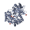
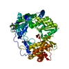
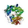
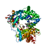
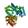
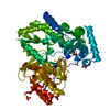
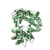
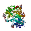
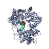
 Hepatitis C virus
 X-RAY DIFFRACTION /
  Authors
 Citation
 Structure visualization
 Downloads & links
 Other downloads
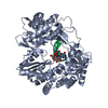

 PDBj
PDBj


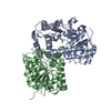
 Assembly
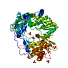


 Mass: 96.063 Da / Num. of mol.: 7 / Source method: obtained synthetically / Formula: SO4
 Mass: 62.068 Da / Num. of mol.: 1 / Source method: obtained synthetically / Formula: C2H6O2
 Mass: 18.015 Da / Num. of mol.: 169 / Source method: isolated from a natural source / Formula: H2O
 Sample preparation
 / Beamline: 5.0.3 / Wavelength: 0.9765 / Wavelength: 0.9765 Å
 Processing