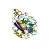
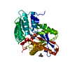
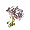
- PDB-4o56: Structure of PLK1 in complex with peptide -
+
データを開く
IDまたはキーワード:
読み込み中...
-
基本情報
登録情報
データベース: PDB / ID: 4o56
タイトル
Structure of PLK1 in complex with peptide
要素
Serine/threonine-protein kinase PLK1
synthetic peptide
キーワード
TRANSFERASE/PEPTIDE / PLK1 Polo Box Domain / Polo box domain / Kinase domain / TRANSFERASE-PEPTIDE complex
機能・相同性
機能・相同性情報
Mitotic Telophase/Cytokinesis / regulation of protein localization to cell cortex / Mitotic Metaphase/Anaphase Transition / synaptonemal complex disassembly / Golgi inheritance / regulation of protein binding / Activation of NIMA Kinases NEK9, NEK6, NEK7 / nuclear membrane disassembly / homologous chromosome segregation / polo kinase ...Mitotic Telophase/Cytokinesis / regulation of protein localization to cell cortex / Mitotic Metaphase/Anaphase Transition / synaptonemal complex disassembly / Golgi inheritance / regulation of protein binding / Activation of NIMA Kinases NEK9, NEK6, NEK7 / nuclear membrane disassembly / homologous chromosome segregation / polo kinase / mitotic nuclear membrane disassembly / Phosphorylation of Emi1 / protein localization to nuclear envelope / metaphase/anaphase transition of mitotic cell cycle / double-strand break repair via alternative nonhomologous end joining / synaptonemal complex / female meiosis chromosome segregation / anaphase-promoting complex binding / Phosphorylation of the APC/C / outer kinetochore / negative regulation of cyclin-dependent protein serine/threonine kinase activity / regulation of mitotic spindle assembly / positive regulation of ubiquitin protein ligase activity / microtubule bundle formation / mitotic chromosome condensation / Polo-like kinase mediated events / Golgi Cisternae Pericentriolar Stack Reorganization / centrosome cycle / regulation of mitotic metaphase/anaphase transition / positive regulation of ubiquitin-protein transferase activity / sister chromatid cohesion / regulation of mitotic cell cycle phase transition / mitotic spindle assembly checkpoint signaling / mitotic spindle pole / regulation of anaphase-promoting complex-dependent catabolic process / mitotic G2 DNA damage checkpoint signaling / establishment of mitotic spindle orientation / positive regulation of proteolysis / mitotic sister chromatid segregation / mitotic cytokinesis / centriolar satellite / negative regulation of double-strand break repair via homologous recombination / spindle midzone / Regulation of MITF-M-dependent genes involved in cell cycle and proliferation / Amplification of signal from unattached kinetochores via a MAD2 inhibitory signal / Cyclin A/B1/B2 associated events during G2/M transition / Mitotic Prometaphase / EML4 and NUDC in mitotic spindle formation / Loss of Nlp from mitotic centrosomes / Loss of proteins required for interphase microtubule organization from the centrosome / Recruitment of mitotic centrosome proteins and complexes / protein localization to chromatin / Recruitment of NuMA to mitotic centrosomes / Anchoring of the basal body to the plasma membrane / regulation of mitotic cell cycle / Resolution of Sister Chromatid Cohesion / centriole / AURKA Activation by TPX2 / mitotic spindle organization / Condensation of Prophase Chromosomes / regulation of cytokinesis / positive regulation of peptidyl-threonine phosphorylation / RHO GTPases Activate Formins / protein destabilization / establishment of protein localization / APC/C:Cdh1 mediated degradation of Cdc20 and other APC/C:Cdh1 targeted proteins in late mitosis/early G1 / kinetochore / spindle pole / positive regulation of protein localization to nucleus / spindle / Separation of Sister Chromatids / The role of GTSE1 in G2/M progression after G2 checkpoint / G2/M transition of mitotic cell cycle / microtubule cytoskeleton / Regulation of PLK1 Activity at G2/M Transition / double-strand break repair / positive regulation of proteasomal ubiquitin-dependent protein catabolic process / mitotic cell cycle / midbody / peptidyl-serine phosphorylation / microtubule binding / regulation of cell cycle / protein kinase activity / protein ubiquitination / protein phosphorylation / protein serine kinase activity / protein serine/threonine kinase activity / centrosome / chromatin / negative regulation of apoptotic process / protein kinase binding / negative regulation of transcription by RNA polymerase II / magnesium ion binding / nucleoplasm / ATP binding / identical protein binding / nucleus / cytosol / cytoplasm 類似検索 - 分子機能
POLO box domain / Polo-like kinase 1, catalytic domain / Second polo-box domain / First polo-box domain / POLO box domain superfamily / POLO box duplicated region / POLO box domain / POLO box domain profile. / Arylsulfatase, C-terminal domain / Serine/threonine-protein kinase, active site ...POLO box domain / Polo-like kinase 1, catalytic domain / Second polo-box domain / First polo-box domain / POLO box domain superfamily / POLO box duplicated region / POLO box domain / POLO box domain profile. / Arylsulfatase, C-terminal domain / Serine/threonine-protein kinase, active site / Serine/Threonine protein kinases active-site signature. / Protein kinase domain / Serine/Threonine protein kinases, catalytic domain / Protein kinase, ATP binding site / Protein kinases ATP-binding region signature. / Protein kinase domain profile. / Protein kinase domain / Protein kinase-like domain superfamily / 2-Layer Sandwich / Alpha Beta 類似検索 - ドメイン・相同性
PHOSPHATE ION / Serine/threonine-protein kinase PLK1 類似検索 - 構成要素
ジャーナル: To be Published タイトル: Integration of Charge-dipole Interaction and Intramolecular Hydrogen Bond in Ligand Design for the Polo-Box Domain of Polo-like Kinase 1 著者: Quan, J.M. / Wang, T. / Shan, H.M.
解像度: 1.8→24.53 Å / Cor.coef. Fo:Fc: 0.968 / Cor.coef. Fo:Fc free: 0.94 / SU B: 4.789 / SU ML: 0.068 / 交差検証法: THROUGHOUT / ESU R: 0.228 / ESU R Free: 0.115 / 立体化学のターゲット値: MAXIMUM LIKELIHOOD / 詳細: HYDROGENS HAVE BEEN ADDED IN THE RIDING POSITIONS
Rfactor
反射数
%反射
Selection details
Rfree
0.2013
1259
5.1 %
RANDOM
Rwork
0.1398
-
-
-
obs
0.14292
23483
99.44 %
-
all
-
24869
-
-
溶媒の処理
イオンプローブ半径: 0.8 Å / 減衰半径: 0.8 Å / VDWプローブ半径: 1.2 Å / 溶媒モデル: MASK
 ムービー
ムービー コントローラー


 データを開く
 基本情報
基本情報 要素
要素 キーワード
キーワード 機能・相同性情報
 Homo sapiens (ヒト)
 X線回折 /
  データ登録者
 引用
引用 構造の表示
構造の表示 ダウンロードとリンク
 その他のダウンロード


 PDBj
PDBj


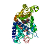
 集合体
集合体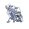


 分子量: 94.971 Da / 分子数: 3 / 由来タイプ: 合成 / 式: PO4
 分子量: 18.015 Da / 分子数: 254 / 由来タイプ: 天然 / 式: H2O
 試料調製
試料調製 解析
解析