 Movie
Movie Controller


+ Open data

- Basic information
| Entry | Database: PDB / ID: 4nzw | ||||||
|---|---|---|---|---|---|---|---|
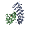
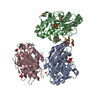
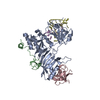
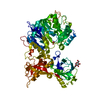
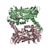
| Title | Crystal Structure of STK25-MO25 Complex | ||||||
 Components Components |
| ||||||
 Keywords Keywords | TRANSFERASE ACTIVATOR/TRANSFERASE / scafolding protein / Ser/Thr protein kinase / TRANSFERASE ACTIVATOR-TRANSFERASE complex | ||||||
| Function / homology |  Function and homology information Function and homology informationintrinsic apoptotic signaling pathway in response to hydrogen peroxide / Golgi localization / negative regulation of potassium ion transmembrane transport / response to thyroid hormone / Golgi reassembly / FAR/SIN/STRIPAK complex / positive regulation of stress-activated MAPK cascade / cellular hypotonic response / establishment of Golgi localization / Energy dependent regulation of mTOR by LKB1-AMPK ...intrinsic apoptotic signaling pathway in response to hydrogen peroxide / Golgi localization / negative regulation of potassium ion transmembrane transport / response to thyroid hormone / Golgi reassembly / FAR/SIN/STRIPAK complex / positive regulation of stress-activated MAPK cascade / cellular hypotonic response / establishment of Golgi localization / Energy dependent regulation of mTOR by LKB1-AMPK / serine/threonine protein kinase complex / positive regulation of axonogenesis / establishment or maintenance of cell polarity / positive regulation of protein serine/threonine kinase activity / protein kinase activator activity / axonogenesis / protein serine/threonine kinase binding / protein serine/threonine kinase activator activity / response to activity / kinase binding / Z disc / protein autophosphorylation / cellular response to oxidative stress / response to oxidative stress / secretory granule lumen / ficolin-1-rich granule lumen / protein phosphorylation / non-specific serine/threonine protein kinase / protein kinase activity / intracellular signal transduction / protein serine kinase activity / protein serine/threonine kinase activity / Neutrophil degranulation / Golgi apparatus / signal transduction / protein homodimerization activity / extracellular exosome / extracellular region / ATP binding / metal ion binding / cytoplasm / cytosol Similarity search - Function | ||||||
| Biological species |  Homo sapiens (human) Homo sapiens (human) | ||||||
| Method |  X-RAY DIFFRACTION / SYNCHROTRON / MOLECULAR REPLACEMENT / Resolution: 3.583 Å X-RAY DIFFRACTION / SYNCHROTRON / MOLECULAR REPLACEMENT / Resolution: 3.583 Å | ||||||
 Authors Authors | Feng, M. / Hao, Q. / Zhou, Z.C. | ||||||
 Citation Citation | Journal: J.Struct.Biol. / Year: 2014 Title: Structural insights into regulatory mechanisms of MO25-mediated kinase activation. Authors: Hao, Q. / Feng, M. / Shi, Z. / Li, C. / Chen, M. / Wang, W. / Zhang, M. / Jiao, S. / Zhou, Z. | ||||||
| History |
|
- Structure visualization
| Structure viewer | Molecule: MolmilJmol/JSmol |
|---|
- Downloads & links
-Download
| PDBx/mmCIF format | 4nzw.cif.gz | 131.9 KB | Display | PDBx/mmCIF format |
|---|---|---|---|---|
| PDB format | pdb4nzw.ent.gz | 100.6 KB | Display | PDB format |
| PDBx/mmJSON format | 4nzw.json.gz | Tree view | PDBx/mmJSON format | |
| Others |  Other downloads Other downloads |
-Validation report
| Summary document | 4nzw_validation.pdf.gz | 672.7 KB | Display | wwPDB validaton report |
|---|---|---|---|---|
| Full document | 4nzw_full_validation.pdf.gz | 684 KB | Display | |
| Data in XML | 4nzw_validation.xml.gz | 22.5 KB | Display | |
| Data in CIF | 4nzw_validation.cif.gz | 29.8 KB | Display | |
| Arichive directory | https://data.pdbj.org/pub/pdb/validation_reports/nz/4nzwftp://data.pdbj.org/pub/pdb/validation_reports/nz/4nzw | HTTPS FTP |
-Related structure data
| Related structure data |  4o27C  1uplS  2xikS S: Starting model for refinement C: citing same article ( |
|---|---|
| Similar structure data |


-Links
 PDBj
PDBj- Assembly
| Deposited unit | 
| ||||||||
|---|---|---|---|---|---|---|---|---|---|
| 1 |
| ||||||||
| Unit cell |
|
-Components
| #1: Protein | Mass: 38665.496 Da / Num. of mol.: 1 / Fragment: UNP RESIDUES 8-334 Source method: isolated from a genetically manipulated source Source: (gene. exp.) Homo sapiens (human) / Gene: CAB39, CGI-66, MO25 / Plasmid: pET28a-HisTEV / Production host:  |
|---|---|
| #2: Protein | Mass: 33704.703 Da / Num. of mol.: 1 / Fragment: kinase domain, UNP RESIDUES 1-293 / Mutation: D158A Source method: isolated from a genetically manipulated source Source: (gene. exp.) Homo sapiens (human) / Gene: SOK1, STK25, YSK1 / Plasmid: pET28a-HisTEV / Production host: References: UniProt: O00506, non-specific serine/threonine protein kinase |
| #3: Chemical | ChemComp-J60 / 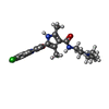  Mass: 414.928 Da / Num. of mol.: 1 / Source method: obtained synthetically / Formula: C22H27ClN4O2 Mass: 414.928 Da / Num. of mol.: 1 / Source method: obtained synthetically / Formula: C22H27ClN4O2 |
-Experimental details
-Experiment
| Experiment | Method: X-RAY DIFFRACTION / Number of used crystals: 1 |
|---|
- Sample preparation
| Crystal | Density Matthews: 2.98 Å3/Da / Density % sol: 58.78 % |
|---|---|
| Crystal grow | Temperature: 277 K / Method: vapor diffusion, sitting drop / pH: 7.5 Details: 0.1M HEPES PH 7.0, 1M SODIUM CITRATE, VAPOR DIFFUSION, SITTING DROP, temperature 277.0K |
-Data collection
| Diffraction | Mean temperature: 100 K |
|---|---|
| Diffraction source | Source: SYNCHROTRON / Site: SSRF  / Beamline: BL17U / Wavelength: 0.9793 Å / Beamline: BL17U / Wavelength: 0.9793 Å |
| Detector | Type: ADSC QUANTUM 315 / Detector: CCD / Date: Jul 3, 2012 |
| Radiation | Monochromator: Si 111 CHANNEL / Protocol: SINGLE WAVELENGTH / Monochromatic (M) / Laue (L): M / Scattering type: x-ray |
| Radiation wavelength | Wavelength: 0.9793 Å / Relative weight: 1 |
| Reflection | Resolution: 3.583→50 Å / Num. all: 10784 / Num. obs: 10753 / % possible obs: 99.7 % / Observed criterion σ(F): 2 / Observed criterion σ(I): 2 / Redundancy: 10.2 % / Rmerge(I) obs: 0.179 / Net I/σ(I): 15.9 |
| Reflection shell | Resolution: 3.6→3.66 Å / Redundancy: 10.3 % / Rmerge(I) obs: 0.963 / Mean I/σ(I) obs: 5.25 / % possible all: 98.45 |
- Processing
| Software |
| ||||||||||||||||||||||||||||||||||||||||||||||||||||||
|---|---|---|---|---|---|---|---|---|---|---|---|---|---|---|---|---|---|---|---|---|---|---|---|---|---|---|---|---|---|---|---|---|---|---|---|---|---|---|---|---|---|---|---|---|---|---|---|---|---|---|---|---|---|---|---|
| Refinement | Method to determine structure: MOLECULAR REPLACEMENT Starting model: 2XIK, 1UPL Resolution: 3.583→40.299 Å / SU ML: 0.37 / σ(F): 1.35 / Phase error: 27.97 / Stereochemistry target values: ML
| ||||||||||||||||||||||||||||||||||||||||||||||||||||||
| Solvent computation | Shrinkage radii: 0.9 Å / VDW probe radii: 1.11 Å / Solvent model: FLAT BULK SOLVENT MODEL | ||||||||||||||||||||||||||||||||||||||||||||||||||||||
| Refinement step | Cycle: LAST / Resolution: 3.583→40.299 Å
| ||||||||||||||||||||||||||||||||||||||||||||||||||||||
| Refine LS restraints |
| ||||||||||||||||||||||||||||||||||||||||||||||||||||||
| LS refinement shell | Refine-ID: X-RAY DIFFRACTION / Total num. of bins used: 8
|
