 Movie
Movie Controller

[English] 日本語
 Yorodumi
- PDB-3mqk: Cbf5-Nop10-Gar1 complex binding with 17mer RNA containing ACA tri... -
+ Open data

- Basic information
| Entry | Database: PDB / ID: 3mqk | ||||||
|---|---|---|---|---|---|---|---|
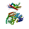
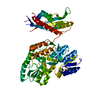
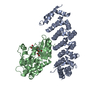
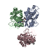
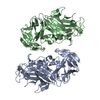
| Title | Cbf5-Nop10-Gar1 complex binding with 17mer RNA containing ACA trinucleotide | ||||||
 Components Components |
| ||||||
 Keywords Keywords | Isomerase/RNA binding protein/rna / Protein-RNA complex / Box H/ACA / pseudouridine synthase / post-transcriptional modification / Isomerase / tRNA processing / RNA-binding / Isomerase-RNA binding protein-rna complex | ||||||
| Function / homology |  Function and homology information Function and homology informationtRNA pseudouridine55 synthase / tRNA pseudouridine(55) synthase activity / pseudouridine synthesis / rRNA pseudouridine synthesis / box H/ACA sno(s)RNA 3'-end processing / tRNA pseudouridine synthesis / snRNA pseudouridine synthesis / mRNA pseudouridine synthesis / snoRNA binding / rRNA processing ...tRNA pseudouridine55 synthase / tRNA pseudouridine(55) synthase activity / pseudouridine synthesis / rRNA pseudouridine synthesis / box H/ACA sno(s)RNA 3'-end processing / tRNA pseudouridine synthesis / snRNA pseudouridine synthesis / mRNA pseudouridine synthesis / snoRNA binding / rRNA processing / ribosome biogenesis / ribonucleoprotein complex / RNA binding Similarity search - Function | ||||||
| Biological species |   Pyrococcus furiosus (archaea) Pyrococcus furiosus (archaea) | ||||||
| Method |  X-RAY DIFFRACTION / SYNCHROTRON / MOLECULAR REPLACEMENT / Resolution: 2.8 Å X-RAY DIFFRACTION / SYNCHROTRON / MOLECULAR REPLACEMENT / Resolution: 2.8 Å | ||||||
 Authors Authors | Zhou, J. / Liang, B. / Li, H. | ||||||
 Citation Citation | Journal: Rna / Year: 2011 Title: Structural and functional evidence of high specificity of Cbf5 for ACA trinucleotide. Authors: Zhou, J. / Liang, B. / Li, H. | ||||||
| History |
|
- Structure visualization
| Structure viewer | Molecule: MolmilJmol/JSmol |
|---|
- Downloads & links
-Download
| PDBx/mmCIF format | 3mqk.cif.gz | 224.4 KB | Display | PDBx/mmCIF format |
|---|---|---|---|---|
| PDB format | pdb3mqk.ent.gz | 178.5 KB | Display | PDB format |
| PDBx/mmJSON format | 3mqk.json.gz | Tree view | PDBx/mmJSON format | |
| Others |  Other downloads Other downloads |
-Validation report
| Arichive directory | https://data.pdbj.org/pub/pdb/validation_reports/mq/3mqkftp://data.pdbj.org/pub/pdb/validation_reports/mq/3mqk | HTTPS FTP |
|---|
-Related structure data
| Related structure data | 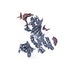 2ey4S S: Starting model for refinement |
|---|---|
| Similar structure data |


-Links
 PDBj
PDBj


- Assembly
| Deposited unit | 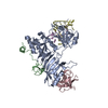
| ||||||||
|---|---|---|---|---|---|---|---|---|---|
| 1 |
| ||||||||
| Unit cell |
|
-Components
| #1: Protein | Mass: 37119.305 Da / Num. of mol.: 1 / Fragment: UNP residues 8-335 Source method: isolated from a genetically manipulated source Source: (gene. exp.) Pyrococcus furiosus (archaea) / Gene: truB, PF1785 / Production host:  References: UniProt: Q7LWY0, Isomerases; Intramolecular transferases; Transferring other groups |
|---|---|
| #2: Protein | Mass: 6205.335 Da / Num. of mol.: 1 / Fragment: UNP residues 4-55 Source method: isolated from a genetically manipulated source Source: (gene. exp.) Pyrococcus furiosus (archaea) / Gene: PF1141 / Production host: |
| #3: Protein | Mass: 8672.303 Da / Num. of mol.: 1 / Fragment: UNP residues 8-82 Source method: isolated from a genetically manipulated source Source: (gene. exp.) Pyrococcus furiosus (archaea) / Gene: PF1791 / Production host: |
| #4: RNA chain | Mass: 4117.501 Da / Num. of mol.: 1 / Source method: obtained synthetically |
| #5: RNA chain | Mass: 2814.759 Da / Num. of mol.: 1 / Source method: obtained synthetically |
| Has protein modification | Y |
-Experimental details
-Experiment
| Experiment | Method: X-RAY DIFFRACTION / Number of used crystals: 1 |
|---|
- Sample preparation
| Crystal | Density Matthews: 3.34 Å3/Da / Density % sol: 63.14 % |
|---|---|
| Crystal grow | Temperature: 303 K / Method: vapor diffusion, hanging drop / pH: 5.6 Details: 120 mM magnesium acetate tetrahydrate, 50 mM MES PH 5.6, 20% 2-Methyl-2,4-pentanediol, VAPOR DIFFUSION, HANGING DROP, temperature 303K |
-Data collection
| Diffraction | Mean temperature: 100 K |
|---|---|
| Diffraction source | Source: SYNCHROTRON / Site: APS  / Beamline: 22-ID / Beamline: 22-ID |
| Detector | Type: MARMOSAIC 300 mm CCD / Detector: CCD / Date: Dec 19, 2007 |
| Radiation | Protocol: SINGLE WAVELENGTH / Monochromatic (M) / Laue (L): M / Scattering type: x-ray |
| Radiation wavelength | Relative weight: 1 |
| Reflection | Resolution: 2.8→100 Å / Num. obs: 20726 / % possible obs: 99.9 % / Redundancy: 20.6 % / Rmerge(I) obs: 0.088 / Net I/σ(I): 48.8 |
| Reflection shell | Resolution: 2.8→2.9 Å / Redundancy: 17.4 % / Rmerge(I) obs: 0.778 / Mean I/σ(I) obs: 3.8 / Num. unique all: 2000 / % possible all: 100 |
- Processing
| Software |
| |||||||||||||||||||||||||||||||||||||||||||||||||||||||||||||||||||||||||||||||||||||||||||||||||||||||||
|---|---|---|---|---|---|---|---|---|---|---|---|---|---|---|---|---|---|---|---|---|---|---|---|---|---|---|---|---|---|---|---|---|---|---|---|---|---|---|---|---|---|---|---|---|---|---|---|---|---|---|---|---|---|---|---|---|---|---|---|---|---|---|---|---|---|---|---|---|---|---|---|---|---|---|---|---|---|---|---|---|---|---|---|---|---|---|---|---|---|---|---|---|---|---|---|---|---|---|---|---|---|---|---|---|---|---|
| Refinement | Method to determine structure: MOLECULAR REPLACEMENT Starting model: PDB entry 2EY4 Resolution: 2.8→33.378 Å / SU ML: 2.55 / σ(F): 0.12 / Stereochemistry target values: ML
| |||||||||||||||||||||||||||||||||||||||||||||||||||||||||||||||||||||||||||||||||||||||||||||||||||||||||
| Solvent computation | Shrinkage radii: 0.9 Å / VDW probe radii: 1.11 Å / Solvent model: FLAT BULK SOLVENT MODEL / Bsol: 51.496 Å2 / ksol: 0.307 e/Å3 | |||||||||||||||||||||||||||||||||||||||||||||||||||||||||||||||||||||||||||||||||||||||||||||||||||||||||
| Displacement parameters |
| |||||||||||||||||||||||||||||||||||||||||||||||||||||||||||||||||||||||||||||||||||||||||||||||||||||||||
| Refinement step | Cycle: LAST / Resolution: 2.8→33.378 Å
| |||||||||||||||||||||||||||||||||||||||||||||||||||||||||||||||||||||||||||||||||||||||||||||||||||||||||
| Refine LS restraints |
| |||||||||||||||||||||||||||||||||||||||||||||||||||||||||||||||||||||||||||||||||||||||||||||||||||||||||
| LS refinement shell |
| |||||||||||||||||||||||||||||||||||||||||||||||||||||||||||||||||||||||||||||||||||||||||||||||||||||||||
| Refinement TLS params. | Method: refined / Origin x: -12.2349 Å / Origin y: 35.566 Å / Origin z: -8.2205 Å
| |||||||||||||||||||||||||||||||||||||||||||||||||||||||||||||||||||||||||||||||||||||||||||||||||||||||||
| Refinement TLS group | Selection details: all |
