[English] 日本語
Yorodumi
- PDB-4nzk: Crystal structure of a DHHW family protein (EUBSIR_00411) from Eu... -

+
Open data


ID or keywords:

Loading...

-
Basic information

Entry
Database: PDB / ID: 4nzk
Title
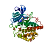
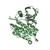
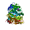
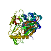
Crystal structure of a DHHW family protein (EUBSIR_00411) from Eubacterium siraeum DSM 15702 at 1.49 A resolution
ComponentsUncharacterized protein
KeywordsSTRUCTURAL GENOMICS / UNKNOWN FUNCTION / DHHW protein / PF14286 family / Joint Center for Structural Genomics / JCSG / Protein Structure Initiative / PSI-BIOLOGY
Function / homologyDHHW protein / DHHW protein / Prokaryotic membrane lipoprotein lipid attachment site profile. / Unknown ligand / AlgX/AlgJ SGNH hydrolase-like domain-containing protein
Function and homology information
Biological speciesEubacterium siraeum (bacteria)
MethodX-RAY DIFFRACTION / SYNCHROTRON / MAD / Resolution: 1.49 Å
AuthorsJoint Center for Structural Genomics (JCSG)
CitationJournal: To be published
Title: Crystal structure of a hypothetical protein (EUBSIR_00411) from Eubacterium siraeum DSM 15702 at 1.49 A resolution
Authors: Joint Center for Structural Genomics (JCSG)
History
DepositionDec 12, 2013Deposition site: RCSB / Processing site: RCSB
Revision 1.0Jan 15, 2014Provider: repository / Type: Initial release
Revision 1.1Dec 24, 2014Group: Structure summary
Revision 1.2Nov 22, 2017Group: Refinement description / Category: software / Item: _software.classification / _software.name
Revision 1.3Jan 24, 2018Group: Database references / Category: citation_author / Item: _citation_author.name
Revision 1.4Feb 1, 2023Group: Database references / Derived calculations
Category: database_2 / struct_conn ...database_2 / struct_conn / struct_ref_seq_dif / struct_site
Item: _database_2.pdbx_DOI / _database_2.pdbx_database_accession ..._database_2.pdbx_DOI / _database_2.pdbx_database_accession / _struct_conn.pdbx_leaving_atom_flag / _struct_ref_seq_dif.details / _struct_site.pdbx_auth_asym_id / _struct_site.pdbx_auth_comp_id / _struct_site.pdbx_auth_seq_id
Revision 1.5Nov 27, 2024Group: Data collection / Structure summary
Category: chem_comp_atom / chem_comp_bond ...chem_comp_atom / chem_comp_bond / pdbx_entry_details / pdbx_modification_feature
Item: _pdbx_entry_details.has_protein_modification

-
Structure visualization

Structure viewerMolecule:
MolmilJmol/JSmol

Downloads & links

-
Assembly

Deposited unit
A: Uncharacterized protein
hetero molecules


Theoretical massNumber of molelcules
Total (without water)39,5515
Polymers39,3651
Non-polymers1864
Water6,612367
1


  • Idetical with deposited unit
  • defined by author&software
TypeNameSymmetry operationNumber
identity operation1_555x,y,z1
MethodPISA
Unit cell
Length a, b, c (Å)102.125, 102.125, 123.311
Angle α, β, γ (deg.)90.000, 90.000, 120.000
Int Tables number178
Space group name H-MP6122
Components on special symmetry positions
IDModelComponents
11A-694-

HOH

21A-740-

HOH

31A-777-

HOH

41A-817-

HOH

-
Components

#1: Protein Uncharacterized protein


Mass: 39365.039 Da / Num. of mol.: 1
Source method: isolated from a genetically manipulated source
Source: (gene. exp.) Eubacterium siraeum (bacteria) / Strain: DSM 15702 / Gene: EUBSIR_00411 / Plasmid: SpeedET / Production host: Escherichia Coli (E. coli) / Strain (production host): PB1 / References: UniProt: B0MKR4
#2: Chemical ChemComp-EDO / 1,2-ETHANEDIOL / ETHYLENE GLYCOL


Mass: 62.068 Da / Num. of mol.: 3 / Source method: obtained synthetically / Formula: C2H6O2
#3: Chemical ChemComp-UNL / UNKNOWN LIGAND


Num. of mol.: 1 / Source method: obtained synthetically
#4: Water ChemComp-HOH / water


Mass: 18.015 Da / Num. of mol.: 367 / Source method: isolated from a natural source / Formula: H2O
Has protein modificationY
Sequence detailsTHIS CONSTRUCT (RESIDUES 33-384) WAS EXPRESSED WITH A PURIFICATION TAG MGSDKIHHHHHHENLYFQG. THE TAG ...THIS CONSTRUCT (RESIDUES 33-384) WAS EXPRESSED WITH A PURIFICATION TAG MGSDKIHHHHHHENLYFQG. THE TAG WAS REMOVED WITH TEV PROTEASE LEAVING ONLY A GLYCINE (0) FOLLOWED BY THE TARGET SEQUENCE.

-
Experimental details

-
Experiment

ExperimentMethod: X-RAY DIFFRACTION / Number of used crystals: 1

-
Sample preparation

CrystalDensity Matthews: 2.36 Å3/Da / Density % sol: 47.83 %
Crystal growTemperature: 277 K / Method: vapor diffusion, sitting drop
Details: 0.20M Sodium Iodide, 20.0% polyethylene glycol 3350, NANODROP, VAPOR DIFFUSION, SITTING DROP, temperature 277K

-
Data collection

DiffractionMean temperature: 100 K
Diffraction sourceSource: SYNCHROTRON / Site: ALS / Beamline: 8.2.1 / Wavelength: 0.918401,0.979346,0.979261
DetectorType: ADSC QUANTUM 315 / Detector: CCD / Date: Oct 17, 2013 / Details: KOHZU: Double Crystal Si(111)
RadiationMonochromator: Double Crystal Si(111) / Protocol: MAD / Monochromatic (M) / Laue (L): M / Scattering type: x-ray
Radiation wavelength
IDWavelength (Å)Relative weight
10.9184011
20.9793461
30.9792611
ReflectionResolution: 1.49→29.481 Å / Num. all: 62386 / Num. obs: 62386 / % possible obs: 100 % / Redundancy: 10.3 % / Rsym value: 0.11 / Net I/σ(I): 12.5
Reflection shell

Rmerge(I) obs: 0.015 / Diffraction-ID: 1

Resolution (Å)Redundancy (%)Mean I/σ(I) obsNum. measured allNum. unique allRsym value% possible all
1.49-1.5310.30.54660245431.482100
1.53-1.5710.30.64594544441.094100
1.57-1.6210.40.84464042770.839100
1.62-1.6710.51.14373041800.647100
1.67-1.7210.51.54253740600.499100
1.72-1.7810.51.84127839400.398100
1.78-1.8510.52.53969237900.297100
1.85-1.9210.43.43841836810.218100
1.92-2.0110.44.33666335160.169100
2.01-2.1110.45.13497733730.137100
2.11-2.2210.363339332280.115100
2.22-2.3610.36.63132130480.104100
2.36-2.5210.26.82938528750.097100
2.52-2.7210.17.12701826880.09100
2.72-2.989.87.32434424910.086100
2.98-3.339.482153622790.078100
3.33-3.851010.52016920250.058100
3.85-4.719.913.71717717330.044100
4.71-6.6610.213.31408013830.045100
6.66-29.4819.19.976058320.04898.9

-
Phasing

PhasingMethod: MAD

-
Processing

Software
NameVersionClassificationNB
MolProbity3beta29model building
PDB_EXTRACT3.1data extraction
SHELXphasing
SHARPphasing
SCALA3.3.20data scaling
REFMAC5.7.0032refinement
MOSFLMdata reduction
SHELXDphasing
RefinementMethod to determine structure: MAD / Resolution: 1.49→29.481 Å / Cor.coef. Fo:Fc: 0.98 / Cor.coef. Fo:Fc free: 0.972 / Occupancy max: 1 / Occupancy min: 0.2 / SU B: 2.017 / SU ML: 0.033 / Cross valid method: THROUGHOUT / σ(F): 0 / ESU R: 0.056 / ESU R Free: 0.053
Stereochemistry target values: MAXIMUM LIKELIHOOD WITH PHASES
Details: 1.HYDROGENS HAVE BEEN ADDED IN THE RIDING POSITIONS. 2.A MET-INHIBITION PROTOCOL WAS USED FOR SELENOMETHIONINE INCORPORATION DURING PROTEIN EXPRESSION. THE OCCUPANCY OF THE SE ATOMS IN THE ...Details: 1.HYDROGENS HAVE BEEN ADDED IN THE RIDING POSITIONS. 2.A MET-INHIBITION PROTOCOL WAS USED FOR SELENOMETHIONINE INCORPORATION DURING PROTEIN EXPRESSION. THE OCCUPANCY OF THE SE ATOMS IN THE MSE RESIDUES WAS REDUCED TO 0.75 FOR THE REDUCED SCATTERING POWER DUE TO PARTIAL S-MET INCORPORATION. 3.1,2-ETHANEDIOL (EDO) FROM THE CRYOPROTECTANT SOLUTION SOLUTION HAS BEEN MODELED IN THE SOLVENT STRUCTURE. 4.N-TERMINAL RESIDUES 34-81 ARE DISORDERED AND ARE EXCLUDED FROM THE MODEL. 5.AN UNIDENTIFIED LIGAND (UNL) HAS BEEN MODELED INTO ELECTRON DENSITY AT THE PUTATIVE ACTIVE SITE BOUND TO CYS365 AND HIS194. THE VICINITY INCLUDES ADDITIONAL UNMODELED ELECTRON DENSITY THAT MAY BE A PORTION OF SOME ENDOGENOUS SUBSTRATE.
RfactorNum. reflection% reflectionSelection details
Rfree0.1577 3144 5 %RANDOM
Rwork0.1247 ---
obs0.1264 62267 99.85 %-
Solvent computationIon probe radii: 0.8 Å / Shrinkage radii: 0.8 Å / VDW probe radii: 1.2 Å / Solvent model: BABINET MODEL WITH MASK
Displacement parametersBiso max: 78.99 Å2 / Biso mean: 19.684 Å2 / Biso min: 5.93 Å2
Baniso -1Baniso -2Baniso -3
1-0.28 Å20.28 Å20 Å2
2--0.28 Å20 Å2
3----0.91 Å2
Refinement stepCycle: LAST / Resolution: 1.49→29.481 Å
ProteinNucleic acidLigandSolventTotal
Num. atoms2389 0 14 367 2770
Refine LS restraints
Refine-IDTypeDev idealDev ideal targetNumber
X-RAY DIFFRACTIONr_bond_refined_d0.0120.022609
X-RAY DIFFRACTIONr_bond_other_d0.0010.022363
X-RAY DIFFRACTIONr_angle_refined_deg1.6531.9473576
X-RAY DIFFRACTIONr_angle_other_deg0.81635468
X-RAY DIFFRACTIONr_dihedral_angle_1_deg5.45350
X-RAY DIFFRACTIONr_dihedral_angle_2_deg32.80924.692130
X-RAY DIFFRACTIONr_dihedral_angle_3_deg11.415390
X-RAY DIFFRACTIONr_dihedral_angle_4_deg12.663158
X-RAY DIFFRACTIONr_chiral_restr0.1010.2387
X-RAY DIFFRACTIONr_gen_planes_refined0.0070.023060
X-RAY DIFFRACTIONr_gen_planes_other0.0010.02638
X-RAY DIFFRACTIONr_mcbond_it2.2083.0541259
X-RAY DIFFRACTIONr_mcbond_other2.2023.0491258
X-RAY DIFFRACTIONr_mcangle_it2.65.7541584
X-RAY DIFFRACTIONr_rigid_bond_restr3.42434972
X-RAY DIFFRACTIONr_sphericity_free27.1215126
X-RAY DIFFRACTIONr_sphericity_bonded9.65155144
LS refinement shellResolution: 1.49→1.529 Å / Total num. of bins used: 20
RfactorNum. reflection% reflection
Rfree0.326 227 -
Rwork0.255 4302 -
all-4529 -
obs--99.74 %

+
About Yorodumi

-
News

-
Feb 9, 2022. New format data for meta-information of EMDB entries

New format data for meta-information of EMDB entries

  • Version 3 of the EMDB header file is now the official format.
  • The previous official version 1.9 will be removed from the archive.

Related info.:EMDB header

External links:wwPDB to switch to version 3 of the EMDB data model

-
Aug 12, 2020. Covid-19 info

Covid-19 info

URL: https://pdbj.org/emnavi/covid19.php

New page: Covid-19 featured information page in EM Navigator.

Related info.:Covid-19 info / Mar 5, 2020. Novel coronavirus structure data

+
Mar 5, 2020. Novel coronavirus structure data

Novel coronavirus structure data

Related info.:Yorodumi Speices / Aug 12, 2020. Covid-19 info

External links:COVID-19 featured content - PDBj / Molecule of the Month (242):Coronavirus Proteases

+
Jan 31, 2019. EMDB accession codes are about to change! (news from PDBe EMDB page)

EMDB accession codes are about to change! (news from PDBe EMDB page)

  • The allocation of 4 digits for EMDB accession codes will soon come to an end. Whilst these codes will remain in use, new EMDB accession codes will include an additional digit and will expand incrementally as the available range of codes is exhausted. The current 4-digit format prefixed with “EMD-” (i.e. EMD-XXXX) will advance to a 5-digit format (i.e. EMD-XXXXX), and so on. It is currently estimated that the 4-digit codes will be depleted around Spring 2019, at which point the 5-digit format will come into force.
  • The EM Navigator/Yorodumi systems omit the EMD- prefix.

Related info.:Q: What is EMD? / ID/Accession-code notation in Yorodumi/EM Navigator

External links:EMDB Accession Codes are Changing Soon! / Contact to PDBj

+
Jul 12, 2017. Major update of PDB

Major update of PDB

  • wwPDB released updated PDB data conforming to the new PDBx/mmCIF dictionary.
  • This is a major update changing the version number from 4 to 5, and with Remediation, in which all the entries are updated.
  • In this update, many items about electron microscopy experimental information are reorganized (e.g. em_software).
  • Now, EM Navigator and Yorodumi are based on the updated data.

External links:wwPDB Remediation / Enriched Model Files Conforming to OneDep Data Standards Now Available in the PDB FTP Archive

-
Yorodumi

Thousand views of thousand structures

  • Yorodumi is a browser for structure data from EMDB, PDB, SASBDB, etc.
  • This page is also the successor to EM Navigator detail page, and also detail information page/front-end page for Omokage search.
  • The word "yorodu" (or yorozu) is an old Japanese word meaning "ten thousand". "mi" (miru) is to see.

Related info.:EMDB / PDB / SASBDB / Comparison of 3 databanks / Yorodumi Search / Aug 31, 2016. New EM Navigator & Yorodumi / Yorodumi Papers / Jmol/JSmol / Function and homology information / Changes in new EM Navigator and Yorodumi

Read more