Entry Database : PDB / ID : 4ny0Title Crystal structure of FERM domain of human focal adhesion kinase Focal adhesion kinase 1 Keywords / / / / Function / homology Function Domain/homology Component
/ / / / / / / / / / / / / / / / / / / / / / / / / / / / / / / / / / / / / / / / / / / / / / / / / / / / / / / / / / / / / / / / / / / / / / / / / / / / / / / / / / / / / / / / / / / / / / / / / / / / / / / / / / / / / / / / / / / / / / / / / / / / / / / / / / / / / / / / / / / / / Biological species Homo sapiens (human)Method / / / / Resolution : 2.8 Å Authors Walkiewicz, K. / Arold, S.T. Journal : Embo J. / Year : 2014Title : FAK dimerization controls its kinase-dependent functions at focal adhesions.Authors : Brami-Cherrier, K. / Gervasi, N. / Arsenieva, D. / Walkiewicz, K. / Boutterin, M.C. / Ortega, A. / Leonard, P.G. / Seantier, B. / Gasmi, L. / Bouceba, T. / Kadare, G. / Girault, J.A. / Arold, S.T. History Deposition Dec 10, 2013 Deposition site / Processing site Revision 1.0 Mar 12, 2014 Provider / Type Revision 1.1 Mar 20, 2024 Group / Database referencesCategory chem_comp_atom / chem_comp_bond ... chem_comp_atom / chem_comp_bond / database_2 / struct_ref_seq_dif Item / _database_2.pdbx_database_accession / _struct_ref_seq_dif.details
Show all Show less
 Movie
Movie Controller


 Open data
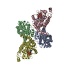
 Basic information
 Components
 Keywords
 Function and homology information
 Homo sapiens (human)
 X-RAY DIFFRACTION /
  Authors
 Citation
 Structure visualization
 Downloads & links
 Other downloads
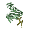


 PDBj
PDBj


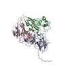
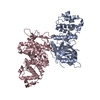
 Assembly
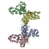


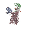
 Sample preparation
 / Beamline: 8.3.1 / Wavelength: 1.115869 Å
 Processing