 Movie
Movie Controller

[English] 日本語
 Yorodumi
- PDB-4nxj: Crystal Structure of PF3D7_1475600, a bromodomain from Plasmodium... -
+ Open data

- Basic information
| Entry | Database: PDB / ID: 4nxj | ||||||
|---|---|---|---|---|---|---|---|
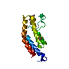
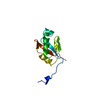
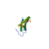
| Title | Crystal Structure of PF3D7_1475600, a bromodomain from Plasmodium Falciparum | ||||||
 Components Components | Bromodomain protein | ||||||
 Keywords Keywords | DNA BINDING PROTEIN / Structural Genomics / Structural Genomics Consortium / SGC / bromodomain | ||||||
| Function / homology |  Function and homology information Function and homology information: / Bromodomain-like / Histone Acetyltransferase; Chain A / Bromodomain / bromo domain / Bromodomain / Bromodomain (BrD) profile. / Bromodomain-like superfamily / Up-down Bundle / Mainly Alpha Similarity search - Domain/homology | ||||||
| Biological species |  | ||||||
| Method |  X-RAY DIFFRACTION / MOLECULAR REPLACEMENT / Resolution: 2.18 Å X-RAY DIFFRACTION / MOLECULAR REPLACEMENT / Resolution: 2.18 Å | ||||||
 Authors Authors | Wernimont, A.K. / Loppnau, P. / Knapp, S. / Fonseca, M. / Brennan, P.E. / Dong, A. / Walker, J.R. / Arrowsmith, C.H. / Edwards, A.M. / Bountra, C. ...Wernimont, A.K. / Loppnau, P. / Knapp, S. / Fonseca, M. / Brennan, P.E. / Dong, A. / Walker, J.R. / Arrowsmith, C.H. / Edwards, A.M. / Bountra, C. / Hui, R. / Hutchinson, A. / Structural Genomics Consortium (SGC) | ||||||
 Citation Citation | Journal: TO BE PUBLISHED Title: Crystal Structure of PF3D7_1475600, a bromodomain from Plasmodium Falciparum Authors: Wernimont, A.K. / Loppnau, P. / Knapp, S. / Fonseca, M. / Brennan, P.E. / Dong, A. / Walker, J.R. / Arrowsmith, C.H. / Edwards, A.M. / Bountra, C. / Hui, R. / Hutchinson, A. | ||||||
| History |
|
- Structure visualization
| Structure viewer | Molecule: MolmilJmol/JSmol |
|---|
- Downloads & links
-Download
| PDBx/mmCIF format | 4nxj.cif.gz | 161.6 KB | Display | PDBx/mmCIF format |
|---|---|---|---|---|
| PDB format | pdb4nxj.ent.gz | 129.8 KB | Display | PDB format |
| PDBx/mmJSON format | 4nxj.json.gz | Tree view | PDBx/mmJSON format | |
| Others |  Other downloads Other downloads |
-Validation report
| Arichive directory | https://data.pdbj.org/pub/pdb/validation_reports/nx/4nxjftp://data.pdbj.org/pub/pdb/validation_reports/nx/4nxj | HTTPS FTP |
|---|
-Related structure data
| Related structure data |  3g0lS S: Starting model for refinement |
|---|---|
| Similar structure data |


-Links
 PDBj
PDBj- Assembly
| Deposited unit | 
| ||||||||
|---|---|---|---|---|---|---|---|---|---|
| 1 | 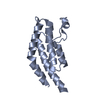
| ||||||||
| 2 | 
| ||||||||
| 3 | 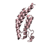
| ||||||||
| Unit cell |
|
-Components
| #1: Protein | Mass: 14432.708 Da / Num. of mol.: 3 / Fragment: UNP residues 8-125 Source method: isolated from a genetically manipulated source Source: (gene. exp.) Strain: 3D7 / Gene: PF14_0724 / Production host:  #2: Water | ChemComp-HOH / |  Mass: 18.015 Da / Num. of mol.: 154 / Source method: isolated from a natural source / Formula: H2O Mass: 18.015 Da / Num. of mol.: 154 / Source method: isolated from a natural source / Formula: H2O |
|---|
-Experimental details
-Experiment
| Experiment | Method: X-RAY DIFFRACTION / Number of used crystals: 1 |
|---|
- Sample preparation
| Crystal | Density Matthews: 2.11 Å3/Da / Density % sol: 41.76 % |
|---|---|
| Crystal grow | Temperature: 293 K / pH: 7.5 Details: 25% PEG3350, 0.3 M KAcetate, pH 7.5, microbatch, temperature 293K |
-Data collection
| Diffraction | Mean temperature: 100 K | |||||||||||||||||||||
|---|---|---|---|---|---|---|---|---|---|---|---|---|---|---|---|---|---|---|---|---|---|---|
| Diffraction source | Source: ROTATING ANODE / Type: RIGAKU FR-E+ SUPERBRIGHT / Wavelength: 1.54178 Å | |||||||||||||||||||||
| Detector | Type: RIGAKU SATURN A200 / Detector: CCD / Date: Sep 27, 2013 | |||||||||||||||||||||
| Radiation | Protocol: SINGLE WAVELENGTH / Monochromatic (M) / Laue (L): M / Scattering type: x-ray | |||||||||||||||||||||
| Radiation wavelength | Wavelength: 1.54178 Å / Relative weight: 1 | |||||||||||||||||||||
| Reflection | Resolution: 2.18→35.75 Å / Num. all: 19612 / Num. obs: 18730 / % possible obs: 95.5 % / Redundancy: 1.7 % / Biso Wilson estimate: 36.35 Å2 / Rmerge(I) obs: 0.081 / Net I/σ(I): 11.1 | |||||||||||||||||||||
| Reflection shell | Diffraction-ID: 1
|
- Processing
| Software |
| ||||||||||||||||||||||||||||||||||||||||||||||||||||||||||||||||||||||||||||||||||||||||||||||||||||||||||||
|---|---|---|---|---|---|---|---|---|---|---|---|---|---|---|---|---|---|---|---|---|---|---|---|---|---|---|---|---|---|---|---|---|---|---|---|---|---|---|---|---|---|---|---|---|---|---|---|---|---|---|---|---|---|---|---|---|---|---|---|---|---|---|---|---|---|---|---|---|---|---|---|---|---|---|---|---|---|---|---|---|---|---|---|---|---|---|---|---|---|---|---|---|---|---|---|---|---|---|---|---|---|---|---|---|---|---|---|---|---|
| Refinement | Method to determine structure: MOLECULAR REPLACEMENT Starting model: FFAS03 model of 3g0l Resolution: 2.18→29.89 Å / Cor.coef. Fo:Fc: 0.92 / Cor.coef. Fo:Fc free: 0.8693 / Occupancy max: 1 / Occupancy min: 0.2 / SU R Cruickshank DPI: 0.319 / Cross valid method: THROUGHOUT / σ(F): 0 / Stereochemistry target values: Engh & Huber
| ||||||||||||||||||||||||||||||||||||||||||||||||||||||||||||||||||||||||||||||||||||||||||||||||||||||||||||
| Displacement parameters | Biso max: 118.51 Å2 / Biso mean: 36.5454 Å2 / Biso min: 7.93 Å2
| ||||||||||||||||||||||||||||||||||||||||||||||||||||||||||||||||||||||||||||||||||||||||||||||||||||||||||||
| Refine analyze | Luzzati coordinate error obs: 0.337 Å | ||||||||||||||||||||||||||||||||||||||||||||||||||||||||||||||||||||||||||||||||||||||||||||||||||||||||||||
| Refinement step | Cycle: LAST / Resolution: 2.18→29.89 Å
| ||||||||||||||||||||||||||||||||||||||||||||||||||||||||||||||||||||||||||||||||||||||||||||||||||||||||||||
| Refine LS restraints |
| ||||||||||||||||||||||||||||||||||||||||||||||||||||||||||||||||||||||||||||||||||||||||||||||||||||||||||||
| LS refinement shell | Resolution: 2.18→2.31 Å / Total num. of bins used: 9
| ||||||||||||||||||||||||||||||||||||||||||||||||||||||||||||||||||||||||||||||||||||||||||||||||||||||||||||
| Refinement TLS params. | Method: refined / Refine-ID: X-RAY DIFFRACTION
| ||||||||||||||||||||||||||||||||||||||||||||||||||||||||||||||||||||||||||||||||||||||||||||||||||||||||||||
| Refinement TLS group |
|
