 Movie
Movie Controller


+ Open data

- Basic information
| Entry | Database: PDB / ID: 4nv5 | ||||||
|---|---|---|---|---|---|---|---|
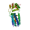
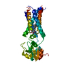
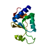
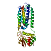
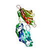
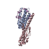
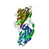
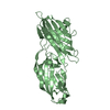
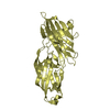
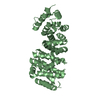
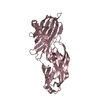
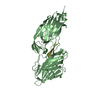
| Title | C50A mutant of Synechococcus VKOR, C2 crystal form (dehydrated) | ||||||
 Components Components | VKORC1/thioredoxin domain protein | ||||||
 Keywords Keywords | OXIDOREDUCTASE / four helix bundle / thioredoxin-like protein / Membrane | ||||||
| Function / homology |  Function and homology information Function and homology informationOxidoreductases; Acting on CH or CH2 groups; With a disulfide as acceptor / quinone binding / oxidoreductase activity / membrane Similarity search - Function | ||||||
| Biological species |  Synechococcus sp. (bacteria) Synechococcus sp. (bacteria) | ||||||
| Method |  X-RAY DIFFRACTION / SYNCHROTRON / MOLECULAR REPLACEMENT / Resolution: 2.79 Å X-RAY DIFFRACTION / SYNCHROTRON / MOLECULAR REPLACEMENT / Resolution: 2.79 Å | ||||||
 Authors Authors | Liu, S. / Cheng, W. / Fowle Grider, R. / Shen, G. / Li, W. | ||||||
 Citation Citation | Journal: Nat Commun / Year: 2014 Title: Structures of an intramembrane vitamin K epoxide reductase homolog reveal control mechanisms for electron transfer. Authors: Liu, S. / Cheng, W. / Fowle Grider, R. / Shen, G. / Li, W. | ||||||
| History |
|
- Structure visualization
| Structure viewer | Molecule: MolmilJmol/JSmol |
|---|
- Downloads & links
-Download
| PDBx/mmCIF format | 4nv5.cif.gz | 210.6 KB | Display | PDBx/mmCIF format |
|---|---|---|---|---|
| PDB format | pdb4nv5.ent.gz | 171.3 KB | Display | PDB format |
| PDBx/mmJSON format | 4nv5.json.gz | Tree view | PDBx/mmJSON format | |
| Others |  Other downloads Other downloads |
-Validation report
| Arichive directory | https://data.pdbj.org/pub/pdb/validation_reports/nv/4nv5ftp://data.pdbj.org/pub/pdb/validation_reports/nv/4nv5 | HTTPS FTP |
|---|
-Related structure data


-Links
 PDBj
PDBj- Assembly
| Deposited unit | 
| ||||||||||||
|---|---|---|---|---|---|---|---|---|---|---|---|---|---|
| 1 | 
| ||||||||||||
| 2 | 
| ||||||||||||
| Unit cell |
| ||||||||||||
| Noncrystallographic symmetry (NCS) | NCS oper:
|
-Components
| #1: Protein | Mass: 31652.775 Da / Num. of mol.: 2 / Mutation: C50A Source method: isolated from a genetically manipulated source Source: (gene. exp.) Synechococcus sp. (bacteria) / Strain: JA-2-3B'a(2-13) / Gene: CYB_2278 / Plasmid: PET20b / Production host: #2: Chemical |   Mass: 863.343 Da / Num. of mol.: 2 / Source method: obtained synthetically / Formula: C59H90O4 Mass: 863.343 Da / Num. of mol.: 2 / Source method: obtained synthetically / Formula: C59H90O4Has protein modification | Y | |
|---|
-Experimental details
-Experiment
| Experiment | Method: X-RAY DIFFRACTION / Number of used crystals: 1 |
|---|
- Sample preparation
| Crystal | Density Matthews: 4.5 Å3/Da / Density % sol: 72.64 % |
|---|---|
| Crystal grow | Temperature: 295 K / Method: vapor diffusion, sitting drop / pH: 5.5 Details: 12% PEG3350, 0.1M sodium cacodylate, pH 5.5, VAPOR DIFFUSION, SITTING DROP, temperature 295K |
-Data collection
| Diffraction | Mean temperature: 77 K | |||||||||||||||
|---|---|---|---|---|---|---|---|---|---|---|---|---|---|---|---|---|
| Diffraction source | Source: SYNCHROTRON / Site: APS  / Beamline: 24-ID-E / Wavelength: 0.97918 / Beamline: 24-ID-E / Wavelength: 0.97918 | |||||||||||||||
| Detector | Type: ADSC QUANTUM 315 / Detector: CCD / Date: Apr 6, 2012 | |||||||||||||||
| Radiation | Monochromator: Si(111) / Protocol: SINGLE WAVELENGTH / Monochromatic (M) / Laue (L): M / Scattering type: x-ray | |||||||||||||||
| Radiation wavelength | Wavelength: 0.97918 Å / Relative weight: 1 | |||||||||||||||
| Reflection twin |
| |||||||||||||||
| Reflection | Resolution: 2.79→50 Å / Num. all: 27621 / Num. obs: 27546 / % possible obs: 99.8 % / Observed criterion σ(F): 1.7 / Observed criterion σ(I): 1.7 / Redundancy: 3.6 % / Rmerge(I) obs: 0.078 / Net I/σ(I): 15.2 | |||||||||||||||
| Reflection shell | Resolution: 2.79→2.9 Å / Redundancy: 3.8 % / Rmerge(I) obs: 0.762 / Mean I/σ(I) obs: 1.7 / % possible all: 100 |
- Processing
| Software |
| ||||||||||||||||||||||||||||||||||||||||||||||||||||||||||||||||||||||||||||||||||||||||||||||||||||||||||||||||||||||||||||||||||||||||||||||||||||||||||||||||||||||||||||||||||||||
|---|---|---|---|---|---|---|---|---|---|---|---|---|---|---|---|---|---|---|---|---|---|---|---|---|---|---|---|---|---|---|---|---|---|---|---|---|---|---|---|---|---|---|---|---|---|---|---|---|---|---|---|---|---|---|---|---|---|---|---|---|---|---|---|---|---|---|---|---|---|---|---|---|---|---|---|---|---|---|---|---|---|---|---|---|---|---|---|---|---|---|---|---|---|---|---|---|---|---|---|---|---|---|---|---|---|---|---|---|---|---|---|---|---|---|---|---|---|---|---|---|---|---|---|---|---|---|---|---|---|---|---|---|---|---|---|---|---|---|---|---|---|---|---|---|---|---|---|---|---|---|---|---|---|---|---|---|---|---|---|---|---|---|---|---|---|---|---|---|---|---|---|---|---|---|---|---|---|---|---|---|---|---|---|
| Refinement | Method to determine structure: MOLECULAR REPLACEMENT / Resolution: 2.79→37.8 Å / Cor.coef. Fo:Fc: 0.915 / Cor.coef. Fo:Fc free: 0.899 / SU B: 18.712 / SU ML: 0.188 / Cross valid method: THROUGHOUT / ESU R: 0.094 / ESU R Free: 0.059 / Stereochemistry target values: MAXIMUM LIKELIHOOD / Details: HYDROGENS HAVE BEEN USED IF PRESENT IN THE INPUT
| ||||||||||||||||||||||||||||||||||||||||||||||||||||||||||||||||||||||||||||||||||||||||||||||||||||||||||||||||||||||||||||||||||||||||||||||||||||||||||||||||||||||||||||||||||||||
| Solvent computation | Ion probe radii: 0.8 Å / Shrinkage radii: 0.8 Å / VDW probe radii: 1.2 Å / Solvent model: MASK | ||||||||||||||||||||||||||||||||||||||||||||||||||||||||||||||||||||||||||||||||||||||||||||||||||||||||||||||||||||||||||||||||||||||||||||||||||||||||||||||||||||||||||||||||||||||
| Displacement parameters | Biso mean: 77.599 Å2
| ||||||||||||||||||||||||||||||||||||||||||||||||||||||||||||||||||||||||||||||||||||||||||||||||||||||||||||||||||||||||||||||||||||||||||||||||||||||||||||||||||||||||||||||||||||||
| Refinement step | Cycle: LAST / Resolution: 2.79→37.8 Å
| ||||||||||||||||||||||||||||||||||||||||||||||||||||||||||||||||||||||||||||||||||||||||||||||||||||||||||||||||||||||||||||||||||||||||||||||||||||||||||||||||||||||||||||||||||||||
| Refine LS restraints |
|