 Movie
Movie Controller

[English] 日本語
 Yorodumi
- PDB-5cfa: Crystal structures of Bbp from Staphylococcus aureus with peptide... -
+ Open data

- Basic information
| Entry | Database: PDB / ID: 5cfa | ||||||
|---|---|---|---|---|---|---|---|
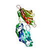
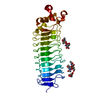
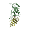
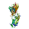
| Title | Crystal structures of Bbp from Staphylococcus aureus with peptide ligand | ||||||
 Components Components |
| ||||||
 Keywords Keywords | PROTEIN BINDING/PEPTIDE / Bbp / Fibrinogen / Sdr / MSCRAMM / Staphylococcus aureus / PROTEIN BINDING-PEPTIDE complex | ||||||
| Function / homology |  Function and homology information Function and homology informationblood coagulation, common pathway / induction of bacterial agglutination / fibrinogen complex / Regulation of TLR by endogenous ligand / platelet alpha granule / MyD88 deficiency (TLR2/4) / positive regulation of heterotypic cell-cell adhesion / IRAK4 deficiency (TLR2/4) / plasminogen activation / MyD88:MAL(TIRAP) cascade initiated on plasma membrane ...blood coagulation, common pathway / induction of bacterial agglutination / fibrinogen complex / Regulation of TLR by endogenous ligand / platelet alpha granule / MyD88 deficiency (TLR2/4) / positive regulation of heterotypic cell-cell adhesion / IRAK4 deficiency (TLR2/4) / plasminogen activation / MyD88:MAL(TIRAP) cascade initiated on plasma membrane / extracellular matrix structural constituent / p130Cas linkage to MAPK signaling for integrins / positive regulation of peptide hormone secretion / GRB2:SOS provides linkage to MAPK signaling for Integrins / positive regulation of exocytosis / blood coagulation, fibrin clot formation / protein polymerization / positive regulation of vasoconstriction / Integrin cell surface interactions / : / negative regulation of endothelial cell apoptotic process / negative regulation of extrinsic apoptotic signaling pathway via death domain receptors / fibrinolysis / Integrin signaling / positive regulation of substrate adhesion-dependent cell spreading / platelet alpha granule lumen / cell-matrix adhesion / positive regulation of protein secretion / Post-translational protein phosphorylation / Signaling by high-kinase activity BRAF mutants / MAP2K and MAPK activation / response to calcium ion / platelet aggregation / Regulation of Insulin-like Growth Factor (IGF) transport and uptake by Insulin-like Growth Factor Binding Proteins (IGFBPs) / Signaling by RAF1 mutants / Signaling by moderate kinase activity BRAF mutants / Paradoxical activation of RAF signaling by kinase inactive BRAF / Signaling downstream of RAS mutants / Signaling by BRAF and RAF1 fusions / Platelet degranulation / extracellular vesicle / extracellular matrix / ER-Phagosome pathway / protein-containing complex assembly / blood microparticle / adaptive immune response / protein-macromolecule adaptor activity / positive regulation of ERK1 and ERK2 cascade / cell adhesion / endoplasmic reticulum lumen / Amyloid fiber formation / signaling receptor binding / external side of plasma membrane / innate immune response / structural molecule activity / cell surface / endoplasmic reticulum / : / extracellular exosome / extracellular region / metal ion binding / plasma membrane Similarity search - Function | ||||||
| Biological species |   Staphylococcus aureus (bacteria) Staphylococcus aureus (bacteria) Homo sapiens (human) Homo sapiens (human) | ||||||
| Method |  X-RAY DIFFRACTION / SYNCHROTRON / MOLECULAR REPLACEMENT / Resolution: 1.45 Å X-RAY DIFFRACTION / SYNCHROTRON / MOLECULAR REPLACEMENT / Resolution: 1.45 Å | ||||||
 Authors Authors | Yu, Y. / Zhang, X.Y. / Gu, J.K. | ||||||
 Citation Citation | Journal: Protein Cell / Year: 2015 Title: Crystal structures of Bbp from Staphylococcus aureus reveal the ligand binding mechanism with Fibrinogen alpha Authors: Zhang, X.Y. / Wu, M. / Zhuo, W. / Gu, J.K. / Zhang, S.S. / Ge, J.P. / Yang, M.J. | ||||||
| History |
|
- Structure visualization
| Structure viewer | Molecule: MolmilJmol/JSmol |
|---|
- Downloads & links
-Download
| PDBx/mmCIF format | 5cfa.cif.gz | 169.9 KB | Display | PDBx/mmCIF format |
|---|---|---|---|---|
| PDB format | pdb5cfa.ent.gz | 130.3 KB | Display | PDB format |
| PDBx/mmJSON format | 5cfa.json.gz | Tree view | PDBx/mmJSON format | |
| Others |  Other downloads Other downloads |
-Validation report
| Arichive directory | https://data.pdbj.org/pub/pdb/validation_reports/cf/5cfaftp://data.pdbj.org/pub/pdb/validation_reports/cf/5cfa | HTTPS FTP |
|---|
-Related structure data
| Related structure data |  5cf3SC S: Starting model for refinement C: citing same article ( |
|---|---|
| Similar structure data |


-Links
 PDBj
PDBj


- Assembly
| Deposited unit | 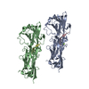
| ||||||||
|---|---|---|---|---|---|---|---|---|---|
| 1 | 
| ||||||||
| 2 | 
| ||||||||
| Unit cell |
|
-Components
| #1: Protein | Mass: 36500.008 Da / Num. of mol.: 2 / Fragment: UNP residues 272-598 Source method: isolated from a genetically manipulated source Source: (gene. exp.) Staphylococcus aureus (bacteria) / Gene: bbp / Production host: #2: Protein/peptide | Mass: 1666.704 Da / Num. of mol.: 2 / Source method: obtained synthetically / Source: (synth.) Homo sapiens (human) / References: UniProt: P02671#3: Chemical | ChemComp-MG / |   Mass: 24.305 Da / Num. of mol.: 1 / Source method: obtained synthetically / Formula: Mg Mass: 24.305 Da / Num. of mol.: 1 / Source method: obtained synthetically / Formula: Mg#4: Water | ChemComp-HOH / |  Mass: 18.015 Da / Num. of mol.: 1026 / Source method: isolated from a natural source / Formula: H2O Mass: 18.015 Da / Num. of mol.: 1026 / Source method: isolated from a natural source / Formula: H2O |
|---|
-Experimental details
-Experiment
| Experiment | Method: X-RAY DIFFRACTION / Number of used crystals: 1 |
|---|
- Sample preparation
| Crystal | Density Matthews: 2.22 Å3/Da / Density % sol: 44.6 % |
|---|---|
| Crystal grow | Temperature: 291.15 K / Method: vapor diffusion, hanging drop Details: peptide was added into the concentrated protein samples at 10:1 ratio and the protein-peptide complex crystals are grown in 0.2 M lithium sulfate, 0.1M Tris-HCl pH8.2, 30% PEG4000 protein ...Details: peptide was added into the concentrated protein samples at 10:1 ratio and the protein-peptide complex crystals are grown in 0.2 M lithium sulfate, 0.1M Tris-HCl pH8.2, 30% PEG4000 protein concentration was 30mg/ml |
-Data collection
| Diffraction | Mean temperature: 100 K | |||||||||||||||||||||||||||||||||||||||||||||||||||||||||||||||||||||||||||||||||||||||||||||||||||
|---|---|---|---|---|---|---|---|---|---|---|---|---|---|---|---|---|---|---|---|---|---|---|---|---|---|---|---|---|---|---|---|---|---|---|---|---|---|---|---|---|---|---|---|---|---|---|---|---|---|---|---|---|---|---|---|---|---|---|---|---|---|---|---|---|---|---|---|---|---|---|---|---|---|---|---|---|---|---|---|---|---|---|---|---|---|---|---|---|---|---|---|---|---|---|---|---|---|---|---|---|
| Diffraction source | Source: SYNCHROTRON / Site: SSRF  / Beamline: BL17U / Wavelength: 0.979 Å / Beamline: BL17U / Wavelength: 0.979 Å | |||||||||||||||||||||||||||||||||||||||||||||||||||||||||||||||||||||||||||||||||||||||||||||||||||
| Detector | Type: ADSC QUANTUM 315r / Detector: CCD / Date: Jun 28, 2014 | |||||||||||||||||||||||||||||||||||||||||||||||||||||||||||||||||||||||||||||||||||||||||||||||||||
| Radiation | Protocol: SINGLE WAVELENGTH / Monochromatic (M) / Laue (L): M / Scattering type: x-ray | |||||||||||||||||||||||||||||||||||||||||||||||||||||||||||||||||||||||||||||||||||||||||||||||||||
| Radiation wavelength | Wavelength: 0.979 Å / Relative weight: 1 | |||||||||||||||||||||||||||||||||||||||||||||||||||||||||||||||||||||||||||||||||||||||||||||||||||
| Reflection | Resolution: 1.45→50 Å / Num. obs: 116230 / % possible obs: 99.2 % / Redundancy: 5.1 % / Biso Wilson estimate: 16.76 Å2 / Rmerge(I) obs: 0.084 / Rpim(I) all: 0.041 / Rrim(I) all: 0.093 / Χ2: 1.408 / Net I/av σ(I): 26.812 / Net I/σ(I): 6.9 / Num. measured all: 591166 | |||||||||||||||||||||||||||||||||||||||||||||||||||||||||||||||||||||||||||||||||||||||||||||||||||
| Reflection shell | Diffraction-ID: 1 / Rejects: _
|
- Processing
| Software |
| |||||||||||||||||||||||||||||||||||||||||||||||||||||||||||||||||||||||||||||||||||||||||||||||||||||||||||||||||||||||||||||||||||||||||||||||||||||||||||||||||||||||||||||||||||||||||||||||||||||||||||||||||||||||||
|---|---|---|---|---|---|---|---|---|---|---|---|---|---|---|---|---|---|---|---|---|---|---|---|---|---|---|---|---|---|---|---|---|---|---|---|---|---|---|---|---|---|---|---|---|---|---|---|---|---|---|---|---|---|---|---|---|---|---|---|---|---|---|---|---|---|---|---|---|---|---|---|---|---|---|---|---|---|---|---|---|---|---|---|---|---|---|---|---|---|---|---|---|---|---|---|---|---|---|---|---|---|---|---|---|---|---|---|---|---|---|---|---|---|---|---|---|---|---|---|---|---|---|---|---|---|---|---|---|---|---|---|---|---|---|---|---|---|---|---|---|---|---|---|---|---|---|---|---|---|---|---|---|---|---|---|---|---|---|---|---|---|---|---|---|---|---|---|---|---|---|---|---|---|---|---|---|---|---|---|---|---|---|---|---|---|---|---|---|---|---|---|---|---|---|---|---|---|---|---|---|---|---|---|---|---|---|---|---|---|---|---|---|---|---|---|---|---|---|
| Refinement | Method to determine structure: MOLECULAR REPLACEMENT Starting model: 5CF3 Resolution: 1.45→36.56 Å / FOM work R set: 0.8246 / SU ML: 0.15 / Cross valid method: FREE R-VALUE / σ(F): 1.34 / Phase error: 24.78 / Stereochemistry target values: ML
| |||||||||||||||||||||||||||||||||||||||||||||||||||||||||||||||||||||||||||||||||||||||||||||||||||||||||||||||||||||||||||||||||||||||||||||||||||||||||||||||||||||||||||||||||||||||||||||||||||||||||||||||||||||||||
| Solvent computation | Shrinkage radii: 0.9 Å / VDW probe radii: 1.11 Å / Solvent model: FLAT BULK SOLVENT MODEL | |||||||||||||||||||||||||||||||||||||||||||||||||||||||||||||||||||||||||||||||||||||||||||||||||||||||||||||||||||||||||||||||||||||||||||||||||||||||||||||||||||||||||||||||||||||||||||||||||||||||||||||||||||||||||
| Displacement parameters | Biso max: 65.31 Å2 / Biso mean: 22.46 Å2 / Biso min: 8 Å2 | |||||||||||||||||||||||||||||||||||||||||||||||||||||||||||||||||||||||||||||||||||||||||||||||||||||||||||||||||||||||||||||||||||||||||||||||||||||||||||||||||||||||||||||||||||||||||||||||||||||||||||||||||||||||||
| Refinement step | Cycle: final / Resolution: 1.45→36.56 Å
| |||||||||||||||||||||||||||||||||||||||||||||||||||||||||||||||||||||||||||||||||||||||||||||||||||||||||||||||||||||||||||||||||||||||||||||||||||||||||||||||||||||||||||||||||||||||||||||||||||||||||||||||||||||||||
| Refine LS restraints |
| |||||||||||||||||||||||||||||||||||||||||||||||||||||||||||||||||||||||||||||||||||||||||||||||||||||||||||||||||||||||||||||||||||||||||||||||||||||||||||||||||||||||||||||||||||||||||||||||||||||||||||||||||||||||||
| LS refinement shell | Refine-ID: X-RAY DIFFRACTION / Total num. of bins used: 30
|
