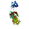
Oxidoreductases; Acting on CH or CH2 groups; With a disulfide as acceptor / quinone binding / oxidoreductase activity / membrane Similarity search - Function
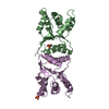
Vitamin K epoxide reductase-like VKOR/LOT1 / VKOR domain / Vitamin K epoxide reductase / VKOR domain superfamily / Vitamin K epoxide reductase family / VKc / de novo design (two linked rop proteins) / Glutaredoxin / Glutaredoxin / Thioredoxin-like superfamily ...Vitamin K epoxide reductase-like VKOR/LOT1 / VKOR domain / Vitamin K epoxide reductase / VKOR domain superfamily / Vitamin K epoxide reductase family / VKc / de novo design (two linked rop proteins) / Glutaredoxin / Glutaredoxin / Thioredoxin-like superfamily / Up-down Bundle / 3-Layer(aba) Sandwich / Mainly Alpha / Alpha Beta Similarity search - Domain/homology
Method to determine structure: MIR / Resolution: 3.6→44.83 Å / Cor.coef. Fo:Fc: 0.861 / Cor.coef. Fo:Fc free: 0.855 / SU B: 44.419 / SU ML: 0.333 / TLS residual ADP flag: LIKELY RESIDUAL / Cross valid method: THROUGHOUT / ESU R Free: 0.581 / Stereochemistry target values: MAXIMUM LIKELIHOOD Details: HYDROGENS HAVE BEEN ADDED IN THE RIDING POSITIONS. Judging from the experimental electron density map, there is density between Cys130 and Cys133, indicative of a disulfide bridge, as well ...Details: HYDROGENS HAVE BEEN ADDED IN THE RIDING POSITIONS. Judging from the experimental electron density map, there is density between Cys130 and Cys133, indicative of a disulfide bridge, as well as density suggesting a covalent bond between the quinone ring of U10 and Cys133. We have not been able to use a refinement program to optimize both bond lengths at the same time
Rfactor
Num. reflection
% reflection
Selection details
Rfree
0.305
855
10 %
RANDOM
Rwork
0.25114
-
-
-
obs
0.25627
7714
99.05 %
-
Solvent computation
Ion probe radii: 0.8 Å / Shrinkage radii: 0.8 Å / VDW probe radii: 1.4 Å / Solvent model: MASK
Displacement parameters
Biso mean: 55.017 Å2
Baniso -1
Baniso -2
Baniso -3
1-
2.74 Å2
1.37 Å2
0 Å2
2-
-
2.74 Å2
0 Å2
3-
-
-
-4.12 Å2
Refinement step
Cycle: LAST / Resolution: 3.6→44.83 Å
Protein
Nucleic acid
Ligand
Solvent
Total
Num. atoms
1963
0
35
0
1998
Refine LS restraints
Refine-ID
Type
Dev ideal
Dev ideal target
Number
X-RAY DIFFRACTION
r_bond_refined_d
0.011
0.022
2051
X-RAY DIFFRACTION
r_bond_other_d
X-RAY DIFFRACTION
r_angle_refined_deg
1.492
1.989
2805
X-RAY DIFFRACTION
r_angle_other_deg
X-RAY DIFFRACTION
r_dihedral_angle_1_deg
7.79
5
256
X-RAY DIFFRACTION
r_dihedral_angle_2_deg
38.577
22.206
68
X-RAY DIFFRACTION
r_dihedral_angle_3_deg
22.001
15
299
X-RAY DIFFRACTION
r_dihedral_angle_4_deg
17.564
15
10
X-RAY DIFFRACTION
r_chiral_restr
0.106
0.2
324
X-RAY DIFFRACTION
r_gen_planes_refined
0.01
0.021
1519
X-RAY DIFFRACTION
r_gen_planes_other
X-RAY DIFFRACTION
r_nbd_refined
X-RAY DIFFRACTION
r_nbd_other
X-RAY DIFFRACTION
r_nbtor_refined
X-RAY DIFFRACTION
r_nbtor_other
X-RAY DIFFRACTION
r_xyhbond_nbd_refined
X-RAY DIFFRACTION
r_xyhbond_nbd_other
X-RAY DIFFRACTION
r_metal_ion_refined
X-RAY DIFFRACTION
r_metal_ion_other
X-RAY DIFFRACTION
r_symmetry_vdw_refined
X-RAY DIFFRACTION
r_symmetry_vdw_other
X-RAY DIFFRACTION
r_symmetry_hbond_refined
X-RAY DIFFRACTION
r_symmetry_hbond_other
X-RAY DIFFRACTION
r_symmetry_metal_ion_refined
X-RAY DIFFRACTION
r_symmetry_metal_ion_other
X-RAY DIFFRACTION
r_mcbond_it
0.355
1.5
1283
X-RAY DIFFRACTION
r_mcbond_other
X-RAY DIFFRACTION
r_mcangle_it
0.692
2
2056
X-RAY DIFFRACTION
r_scbond_it
0.847
3
768
X-RAY DIFFRACTION
r_scangle_it
1.443
4.5
749
X-RAY DIFFRACTION
r_rigid_bond_restr
X-RAY DIFFRACTION
r_sphericity_free
X-RAY DIFFRACTION
r_sphericity_bonded
LS refinement shell
Resolution: 3.6→3.696 Å / Total num. of bins used: 20
Rfactor
Num. reflection
% reflection
Rfree
0.374
67
-
Rwork
0.293
558
-
obs
-
-
96.75 %
Refinement TLS params.
Method: refined / Refine-ID: X-RAY DIFFRACTION
ID
L11 (°2)
L12 (°2)
L13 (°2)
L22 (°2)
L23 (°2)
L33 (°2)
S11 (Å °)
S12 (Å °)
S13 (Å °)
S21 (Å °)
S22 (Å °)
S23 (Å °)
S31 (Å °)
S32 (Å °)
S33 (Å °)
T11 (Å2)
T12 (Å2)
T13 (Å2)
T22 (Å2)
T23 (Å2)
T33 (Å2)
Origin x (Å)
Origin y (Å)
Origin z (Å)
1
46.078
0.5225
-0.4617
15.2841
21.0366
8.2202
0.0983
-2.958
2.3345
0.4986
-0.7731
0.2998
-0.7482
-1.0585
0.6748
2.2956
-1.3821
0.542
2.042
0.5223
1.0831
18.9848
50.9231
12.4686
2
-1.7365
-1.8275
-1.0054
0.7248
2.4627
12.2379
0.3586
0.1278
0.2419
-0.0183
0.6247
-0.6276
2.1193
1.0228
-0.9833
0.8842
-0.3225
-0.2123
1.4523
-0.4603
0.754
40.8754
60.6022
9.7477
3
11.9642
6.7598
-2.241
1.6312
-0.1647
2.5931
-0.5028
1.2689
-0.6467
-0.6585
0.4053
-0.4276
-0.7538
-0.2962
0.0975
1.4677
0.1181
0.1213
1.016
-0.0426
1.518
50.9767
73.6483
16.0345
4
7.4344
2.439
-1.2328
2.0072
1.0314
10.6069
0.1215
0.2352
-0.4932
0.2365
0.0934
0.0945
1.2643
-1.3395
-0.2149
0.5766
-0.6024
-0.112
0.808
0.1032
0.818
32.801
58.6327
22.6571
5
11.334
3.0755
3.806
5.3462
0.2514
6.076
-0.0515
-0.6414
0.3896
0.3344
0.1206
-0.0264
-0.3433
-0.28
-0.0691
0.4952
-0.0531
-0.088
0.4649
-0.0298
1.1823
58.6703
86.0863
23.6554
6
23.6889
-10.9828
3.9082
4.4377
1.5919
16.4774
-1.6816
-0.2654
1.4188
0.7164
0.0549
-0.3331
0.9752
-0.675
1.6267
0.6581
-0.2099
-0.2752
0.6086
-0.0166
1.495
73.3649
85.0948
29.9074
Refinement TLS group
ID
Refine-ID
Refine TLS-ID
Auth asym-ID
Auth seq-ID
1
X-RAY DIFFRACTION
1
A
16 - 20
2
X-RAY DIFFRACTION
2
A
21 - 44
3
X-RAY DIFFRACTION
3
A
45 - 70
4
X-RAY DIFFRACTION
4
A
71 - 177
5
X-RAY DIFFRACTION
5
A
178 - 272
6
X-RAY DIFFRACTION
6
A
273 - 279
+
About Yorodumi
-
News
-
Feb 9, 2022. New format data for meta-information of EMDB entries
New format data for meta-information of EMDB entries
Version 3 of the EMDB header file is now the official format.
The previous official version 1.9 will be removed from the archive.
In the structure databanks used in Yorodumi, some data are registered as the other names, "COVID-19 virus" and "2019-nCoV". Here are the details of the virus and the list of structure data.
Jan 31, 2019. EMDB accession codes are about to change! (news from PDBe EMDB page)
EMDB accession codes are about to change! (news from PDBe EMDB page)
The allocation of 4 digits for EMDB accession codes will soon come to an end. Whilst these codes will remain in use, new EMDB accession codes will include an additional digit and will expand incrementally as the available range of codes is exhausted. The current 4-digit format prefixed with “EMD-” (i.e. EMD-XXXX) will advance to a 5-digit format (i.e. EMD-XXXXX), and so on. It is currently estimated that the 4-digit codes will be depleted around Spring 2019, at which point the 5-digit format will come into force.
The EM Navigator/Yorodumi systems omit the EMD- prefix.
Related info.:Q: What is EMD? / ID/Accession-code notation in Yorodumi/EM Navigator
Yorodumi is a browser for structure data from EMDB, PDB, SASBDB, etc.
This page is also the successor to EM Navigator detail page, and also detail information page/front-end page for Omokage search.
The word "yorodu" (or yorozu) is an old Japanese word meaning "ten thousand". "mi" (miru) is to see.
Related info.:EMDB / PDB / SASBDB / Comparison of 3 databanks / Yorodumi Search / Aug 31, 2016. New EM Navigator & Yorodumi / Yorodumi Papers / Jmol/JSmol / Function and homology information / Changes in new EM Navigator and Yorodumi
 Movie
Movie Controller


 Open data
 Basic information
 Components
 Keywords
 Function and homology information
 Synechococcus sp. (bacteria)
 X-RAY DIFFRACTION /
  Authors
 Citation
 Structure visualization
 Downloads & links
 Other downloads
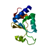


 PDBj
PDBj


 Assembly

 Mass: 863.343 Da / Num. of mol.: 1 / Source method: obtained synthetically / Formula: C59H90O4
 Mass: 200.590 Da / Num. of mol.: 2 / Source method: obtained synthetically / Formula: Hg
 Sample preparation
 / Beamline: 24-ID-C / Wavelength: 1.00718 Å
 Processing