| 登録情報 | データベース: PDB / ID: 4nqq
|
|---|
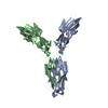
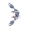
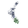
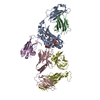
| タイトル | Crystal structure of mouse P-cadherin extracellular domains EC1-EC2 |
|---|
 要素 要素 | Cadherin-3 |
|---|
 キーワード キーワード | CELL ADHESION / Extracellular cadherin (EC) domain / Cell-Cell adhesion |
|---|
| 機能・相同性 |  機能・相同性情報 機能・相同性情報
positive regulation of melanosome transport / negative regulation of timing of catagen / hair cycle process / positive regulation of keratinocyte proliferation / Adherens junctions interactions / positive regulation of melanin biosynthetic process / retina homeostasis / positive regulation of insulin-like growth factor receptor signaling pathway / keratinization / homophilic cell-cell adhesion ...positive regulation of melanosome transport / negative regulation of timing of catagen / hair cycle process / positive regulation of keratinocyte proliferation / Adherens junctions interactions / positive regulation of melanin biosynthetic process / retina homeostasis / positive regulation of insulin-like growth factor receptor signaling pathway / keratinization / homophilic cell-cell adhesion / adherens junction / negative regulation of transforming growth factor beta receptor signaling pathway / cell-cell adhesion / positive regulation of canonical Wnt signaling pathway / response to xenobiotic stimulus / calcium ion binding / positive regulation of gene expression / plasma membrane類似検索 - 分子機能 Cadherin, Y-type LIR-motif / Cadherin, Y-type LIR-motif / Cadherin / Catenin binding domain superfamily / Cadherins / Cadherin conserved site / Cadherin domain signature. / Cadherin repeats. / Cadherin domain / Cadherin-like ...Cadherin, Y-type LIR-motif / Cadherin, Y-type LIR-motif / Cadherin / Catenin binding domain superfamily / Cadherins / Cadherin conserved site / Cadherin domain signature. / Cadherin repeats. / Cadherin domain / Cadherin-like / Cadherins domain profile. / Cadherin-like superfamily / Immunoglobulin-like / Sandwich / Mainly Beta類似検索 - ドメイン・相同性 |
|---|
| 生物種 |   Mus musculus (ハツカネズミ) Mus musculus (ハツカネズミ) |
|---|
| 手法 |  X線回折 / シンクロトロン / 分子置換 / 解像度: 3.2 Å X線回折 / シンクロトロン / 分子置換 / 解像度: 3.2 Å |
|---|
 データ登録者 データ登録者 | Brasch, J. / Shapiro, L. |
|---|
 引用 引用 | ジャーナル: Proc.Natl.Acad.Sci.USA / 年: 2014
タイトル: Structural and energetic determinants of adhesive binding specificity in type I cadherins.
著者: Vendome, J. / Felsovalyi, K. / Song, H. / Yang, Z. / Jin, X. / Brasch, J. / Harrison, O.J. / Ahlsen, G. / Bahna, F. / Kaczynska, A. / Katsamba, P.S. / Edmond, D. / Hubbell, W.L. / Shapiro, L. / Honig, B. |
|---|
| 履歴 | | 登録 | 2013年11月25日 | 登録サイト: RCSB / 処理サイト: RCSB |
|---|
| 改定 1.0 | 2014年9月24日 | Provider: repository / タイプ: Initial release |
|---|
| 改定 1.1 | 2014年10月22日 | Group: Database references |
|---|
| 改定 1.2 | 2017年11月22日 | Group: Refinement description / カテゴリ: software / Item: _software.name |
|---|
| 改定 1.3 | 2023年9月20日 | Group: Data collection / Database references ...Data collection / Database references / Derived calculations / Refinement description
カテゴリ: chem_comp_atom / chem_comp_bond ...chem_comp_atom / chem_comp_bond / database_2 / pdbx_initial_refinement_model / pdbx_struct_conn_angle / struct_conn / struct_site
Item: _database_2.pdbx_DOI / _database_2.pdbx_database_accession ..._database_2.pdbx_DOI / _database_2.pdbx_database_accession / _pdbx_struct_conn_angle.ptnr1_auth_asym_id / _pdbx_struct_conn_angle.ptnr1_auth_comp_id / _pdbx_struct_conn_angle.ptnr1_auth_seq_id / _pdbx_struct_conn_angle.ptnr1_label_asym_id / _pdbx_struct_conn_angle.ptnr1_label_atom_id / _pdbx_struct_conn_angle.ptnr1_label_comp_id / _pdbx_struct_conn_angle.ptnr1_label_seq_id / _pdbx_struct_conn_angle.ptnr2_auth_asym_id / _pdbx_struct_conn_angle.ptnr2_auth_comp_id / _pdbx_struct_conn_angle.ptnr2_auth_seq_id / _pdbx_struct_conn_angle.ptnr2_label_asym_id / _pdbx_struct_conn_angle.ptnr2_label_atom_id / _pdbx_struct_conn_angle.ptnr2_label_comp_id / _pdbx_struct_conn_angle.ptnr3_auth_asym_id / _pdbx_struct_conn_angle.ptnr3_auth_comp_id / _pdbx_struct_conn_angle.ptnr3_auth_seq_id / _pdbx_struct_conn_angle.ptnr3_label_asym_id / _pdbx_struct_conn_angle.ptnr3_label_atom_id / _pdbx_struct_conn_angle.ptnr3_label_comp_id / _pdbx_struct_conn_angle.ptnr3_label_seq_id / _pdbx_struct_conn_angle.value / _struct_conn.pdbx_dist_value / _struct_conn.ptnr1_auth_asym_id / _struct_conn.ptnr1_auth_comp_id / _struct_conn.ptnr1_auth_seq_id / _struct_conn.ptnr1_label_asym_id / _struct_conn.ptnr1_label_atom_id / _struct_conn.ptnr1_label_comp_id / _struct_conn.ptnr1_label_seq_id / _struct_conn.ptnr2_auth_asym_id / _struct_conn.ptnr2_auth_comp_id / _struct_conn.ptnr2_auth_seq_id / _struct_conn.ptnr2_label_asym_id / _struct_conn.ptnr2_label_atom_id / _struct_conn.ptnr2_label_comp_id / _struct_site.pdbx_auth_asym_id / _struct_site.pdbx_auth_comp_id / _struct_site.pdbx_auth_seq_id |
|---|
|
|---|
 ムービー
ムービー コントローラー


 データを開く
 基本情報
基本情報 構造の表示
構造の表示 ダウンロードとリンク
 その他のダウンロード


 PDBj
PDBj
 集合体
集合体


 分子量: 40.078 Da / 分子数: 14 / 由来タイプ: 合成 / 式: Ca
 分子量: 63.546 Da / 分子数: 9 / 由来タイプ: 合成 / 式: Cu
 分子量: 18.015 Da / 分子数: 76 / 由来タイプ: 天然 / 式: H2O
 試料調製
試料調製 / ビームライン: X4C / 波長: 0.9792 Å
 解析
解析