 Movie
Movie Controller


+ Open data

- Basic information
| Entry | Database: PDB / ID: 4npk | ||||||
|---|---|---|---|---|---|---|---|
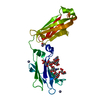
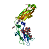
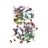
| Title | Extended-Synaptotagmin 2, C2A- and C2B-domains, calcium bound | ||||||
 Components Components | Extended synaptotagmin-2 | ||||||
 Keywords Keywords | MEMBRANE PROTEIN / CALCIUM/PHOSPHOLIPID BINDING PROTEIN / C2 domain / ER to plasma membrane / membrane traffic / protein targeting / Plasma membrane | ||||||
| Function / homology |  Function and homology information Function and homology informationendoplasmic reticulum-plasma membrane tethering / endoplasmic reticulum-plasma membrane contact site / organelle membrane contact site / Glycosphingolipid transport / phosphatidylcholine binding / calcium-dependent phospholipid binding / phosphatidylethanolamine binding / lipid transport / phosphatidylinositol binding / cytoplasmic side of plasma membrane ...endoplasmic reticulum-plasma membrane tethering / endoplasmic reticulum-plasma membrane contact site / organelle membrane contact site / Glycosphingolipid transport / phosphatidylcholine binding / calcium-dependent phospholipid binding / phosphatidylethanolamine binding / lipid transport / phosphatidylinositol binding / cytoplasmic side of plasma membrane / endocytosis / cadherin binding / calcium ion binding / endoplasmic reticulum membrane / identical protein binding / membrane / plasma membrane / cytosol Similarity search - Function | ||||||
| Biological species |  Homo sapiens (human) Homo sapiens (human) | ||||||
| Method |  X-RAY DIFFRACTION / SYNCHROTRON / MOLECULAR REPLACEMENT / Resolution: 2.552 Å X-RAY DIFFRACTION / SYNCHROTRON / MOLECULAR REPLACEMENT / Resolution: 2.552 Å | ||||||
 Authors Authors | Tomchick, D.R. / Rizo, J. / Xu, J. | ||||||
 Citation Citation | Journal: Structure / Year: 2014 Title: Structure and ca(2+)-binding properties of the tandem c2 domains of e-syt2. Authors: Xu, J. / Bacaj, T. / Zhou, A. / Tomchick, D.R. / Sudhof, T.C. / Rizo, J. #1: Journal: Proc.Natl.Acad.Sci.USA / Year: 2007 Title: E-Syts, a family of membranous Ca2+-sensor proteins with multiple C2 domains. Authors: Min, S.W. / Chang, W.P. / Sudhof, T.C. | ||||||
| History |
|
- Structure visualization
| Structure viewer | Molecule: MolmilJmol/JSmol |
|---|
- Downloads & links
-Download
| PDBx/mmCIF format | 4npk.cif.gz | 175.3 KB | Display | PDBx/mmCIF format |
|---|---|---|---|---|
| PDB format | pdb4npk.ent.gz | 141.6 KB | Display | PDB format |
| PDBx/mmJSON format | 4npk.json.gz | Tree view | PDBx/mmJSON format | |
| Others |  Other downloads Other downloads |
-Validation report
| Summary document | 4npk_validation.pdf.gz | 437.6 KB | Display | wwPDB validaton report |
|---|---|---|---|---|
| Full document | 4npk_full_validation.pdf.gz | 440.3 KB | Display | |
| Data in XML | 4npk_validation.xml.gz | 11.4 KB | Display | |
| Data in CIF | 4npk_validation.cif.gz | 14.2 KB | Display | |
| Arichive directory | https://data.pdbj.org/pub/pdb/validation_reports/np/4npkftp://data.pdbj.org/pub/pdb/validation_reports/np/4npk | HTTPS FTP |
-Related structure data
| Related structure data |  4npjSC S: Starting model for refinement C: citing same article ( |
|---|---|
| Similar structure data |


-Links
 PDBj
PDBj


- Assembly
| Deposited unit | 
| ||||||||
|---|---|---|---|---|---|---|---|---|---|
| 1 |
| ||||||||
| Unit cell |
|
-Components
| #1: Protein | Mass: 34265.121 Da / Num. of mol.: 1 / Fragment: C2A and C2B domains / Mutation: A512V Source method: isolated from a genetically manipulated source Source: (gene. exp.) Homo sapiens (human) / Gene: ESYT2, extended-synaptotagmin 2, FAM62B, KIAA1228 / Production host:  | ||
|---|---|---|---|
| #2: Chemical |   Mass: 40.078 Da / Num. of mol.: 3 / Source method: obtained synthetically / Formula: Ca Mass: 40.078 Da / Num. of mol.: 3 / Source method: obtained synthetically / Formula: Ca#3: Water | ChemComp-HOH / |  Mass: 18.015 Da / Num. of mol.: 4 / Source method: isolated from a natural source / Formula: H2O Mass: 18.015 Da / Num. of mol.: 4 / Source method: isolated from a natural source / Formula: H2O |
-Experimental details
-Experiment
| Experiment | Method: X-RAY DIFFRACTION / Number of used crystals: 1 |
|---|
- Sample preparation
| Crystal | Density Matthews: 3.12 Å3/Da / Density % sol: 60.63 % |
|---|---|
| Crystal grow | Temperature: 293 K / Method: vapor diffusion, hanging drop / pH: 8 Details: 6% PEG 8000, 0.1 M Hepes pH 8.0, 0.125 M NaCl, 2.5% glycerol, 0.02 M CaCl2, 0.5 mM TCEP, vapor diffusion, hanging drop, temperature 293K |
-Data collection
| Diffraction | Mean temperature: 100 K |
|---|---|
| Diffraction source | Source: SYNCHROTRON / Site: APS  / Beamline: 19-ID / Wavelength: 0.97912 Å / Beamline: 19-ID / Wavelength: 0.97912 Å |
| Detector | Type: ADSC QUANTUM 315r / Detector: CCD / Date: Oct 18, 2012 / Details: monochromator |
| Radiation | Monochromator: SAGITALLY FOCUSED Si(111) / Protocol: SINGLE WAVELENGTH / Scattering type: x-ray |
| Radiation wavelength | Wavelength: 0.97912 Å / Relative weight: 1 |
| Reflection | Resolution: 2.55→30 Å / Num. all: 12318 / Num. obs: 12318 / % possible obs: 88.7 % / Observed criterion σ(F): 0 / Observed criterion σ(I): 0 / Redundancy: 3.6 % / Biso Wilson estimate: 68.45 Å2 / Rmerge(I) obs: 0.058 / Net I/σ(I): 33.4 |
| Reflection shell | Resolution: 2.55→2.59 Å / Redundancy: 2.4 % / Rmerge(I) obs: 0.276 / Mean I/σ(I) obs: 2.45 / Num. unique all: 263 / % possible all: 38 |
- Processing
| Software |
| ||||||||||||||||||||||||||||||||||||||||||||||||||||||||||||||||||||||||||||||||||||||||||||||||||||||||||||||||||||||||||||||||||||||||||||||||||||||||||||||||||||||||||||||||||||||||||||||||||||||||
|---|---|---|---|---|---|---|---|---|---|---|---|---|---|---|---|---|---|---|---|---|---|---|---|---|---|---|---|---|---|---|---|---|---|---|---|---|---|---|---|---|---|---|---|---|---|---|---|---|---|---|---|---|---|---|---|---|---|---|---|---|---|---|---|---|---|---|---|---|---|---|---|---|---|---|---|---|---|---|---|---|---|---|---|---|---|---|---|---|---|---|---|---|---|---|---|---|---|---|---|---|---|---|---|---|---|---|---|---|---|---|---|---|---|---|---|---|---|---|---|---|---|---|---|---|---|---|---|---|---|---|---|---|---|---|---|---|---|---|---|---|---|---|---|---|---|---|---|---|---|---|---|---|---|---|---|---|---|---|---|---|---|---|---|---|---|---|---|---|---|---|---|---|---|---|---|---|---|---|---|---|---|---|---|---|---|---|---|---|---|---|---|---|---|---|---|---|---|---|---|---|---|
| Refinement | Method to determine structure: MOLECULAR REPLACEMENT Starting model: pdb entry 4NPJ Resolution: 2.552→28.986 Å / Occupancy max: 1 / Occupancy min: 1 / SU ML: 0.44 / Isotropic thermal model: OVERALL / Cross valid method: THROUGHOUT / σ(F): 0 / Phase error: 42.82 / Stereochemistry target values: ML Details: Used reference model restraints. Reference model was
| ||||||||||||||||||||||||||||||||||||||||||||||||||||||||||||||||||||||||||||||||||||||||||||||||||||||||||||||||||||||||||||||||||||||||||||||||||||||||||||||||||||||||||||||||||||||||||||||||||||||||
| Solvent computation | Shrinkage radii: 0.9 Å / VDW probe radii: 1.11 Å / Solvent model: FLAT BULK SOLVENT MODEL | ||||||||||||||||||||||||||||||||||||||||||||||||||||||||||||||||||||||||||||||||||||||||||||||||||||||||||||||||||||||||||||||||||||||||||||||||||||||||||||||||||||||||||||||||||||||||||||||||||||||||
| Displacement parameters | Biso max: 275.75 Å2 / Biso mean: 133.5576 Å2 / Biso min: 69.44 Å2 | ||||||||||||||||||||||||||||||||||||||||||||||||||||||||||||||||||||||||||||||||||||||||||||||||||||||||||||||||||||||||||||||||||||||||||||||||||||||||||||||||||||||||||||||||||||||||||||||||||||||||
| Refine analyze | Luzzati sigma a free: 0.44 Å | ||||||||||||||||||||||||||||||||||||||||||||||||||||||||||||||||||||||||||||||||||||||||||||||||||||||||||||||||||||||||||||||||||||||||||||||||||||||||||||||||||||||||||||||||||||||||||||||||||||||||
| Refinement step | Cycle: LAST / Resolution: 2.552→28.986 Å
| ||||||||||||||||||||||||||||||||||||||||||||||||||||||||||||||||||||||||||||||||||||||||||||||||||||||||||||||||||||||||||||||||||||||||||||||||||||||||||||||||||||||||||||||||||||||||||||||||||||||||
| Refine LS restraints |
| ||||||||||||||||||||||||||||||||||||||||||||||||||||||||||||||||||||||||||||||||||||||||||||||||||||||||||||||||||||||||||||||||||||||||||||||||||||||||||||||||||||||||||||||||||||||||||||||||||||||||
| LS refinement shell | Refine-ID: X-RAY DIFFRACTION / Total num. of bins used: 4
| ||||||||||||||||||||||||||||||||||||||||||||||||||||||||||||||||||||||||||||||||||||||||||||||||||||||||||||||||||||||||||||||||||||||||||||||||||||||||||||||||||||||||||||||||||||||||||||||||||||||||
| Refinement TLS params. | Method: refined / Refine-ID: X-RAY DIFFRACTION
| ||||||||||||||||||||||||||||||||||||||||||||||||||||||||||||||||||||||||||||||||||||||||||||||||||||||||||||||||||||||||||||||||||||||||||||||||||||||||||||||||||||||||||||||||||||||||||||||||||||||||
| Refinement TLS group |
|
