 Movie
Movie Controller

[English] 日本語
 Yorodumi
- PDB-4nng: Structural basis for targeting the ribosomal protein S1 of Mycoba... -
+ Open data

- Basic information
| Entry | Database: PDB / ID: 4nng | ||||||
|---|---|---|---|---|---|---|---|
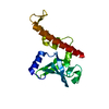
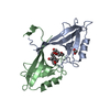
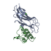
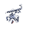
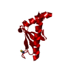
| Title | Structural basis for targeting the ribosomal protein S1 of Mycobacterium tuberculosis by pyrazinamide | ||||||
 Components Components | 30S ribosomal protein S1 | ||||||
 Keywords Keywords | RIBOSOMAL PROTEIN / beta barrel | ||||||
| Function / homology |  Function and homology information Function and homology informationpeptidoglycan-based cell wall / cytosolic small ribosomal subunit / structural constituent of ribosome / translation / mRNA binding / plasma membrane / cytosol Similarity search - Function | ||||||
| Biological species |   Mycobacterium tuberculosis (bacteria) Mycobacterium tuberculosis (bacteria) | ||||||
| Method |  X-RAY DIFFRACTION / SYNCHROTRON / MAD / molecular replacement / Resolution: 2.02 Å X-RAY DIFFRACTION / SYNCHROTRON / MAD / molecular replacement / Resolution: 2.02 Å | ||||||
 Authors Authors | Yang, J. / Liu, Y. / Cai, Q. / Lin, D. | ||||||
 Citation Citation | Journal: Mol.Microbiol. / Year: 2015 Title: Structural basis for targeting the ribosomal protein S1 of Mycobacterium tuberculosis by pyrazinamide. Authors: Yang, J. / Liu, Y. / Bi, J. / Cai, Q. / Liao, X. / Li, W. / Guo, C. / Zhang, Q. / Lin, T. / Zhao, Y. / Wang, H. / Liu, J. / Zhang, X. / Lin, D. | ||||||
| History |
|
- Structure visualization
| Structure viewer | Molecule: MolmilJmol/JSmol |
|---|
- Downloads & links
-Download
| PDBx/mmCIF format | 4nng.cif.gz | 46.8 KB | Display | PDBx/mmCIF format |
|---|---|---|---|---|
| PDB format | pdb4nng.ent.gz | 32.3 KB | Display | PDB format |
| PDBx/mmJSON format | 4nng.json.gz | Tree view | PDBx/mmJSON format | |
| Others |  Other downloads Other downloads |
-Validation report
| Arichive directory | https://data.pdbj.org/pub/pdb/validation_reports/nn/4nngftp://data.pdbj.org/pub/pdb/validation_reports/nn/4nng | HTTPS FTP |
|---|
-Related structure data


-Links
 PDBj
PDBj


- Assembly
| Deposited unit | 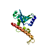
| ||||||||
|---|---|---|---|---|---|---|---|---|---|
| 1 |
| ||||||||
| Unit cell |
|
-Components
| #1: Protein | Mass: 18344.479 Da / Num. of mol.: 1 / Fragment: UNP RESIDUES 285-435 / Mutation: P298A Source method: isolated from a genetically manipulated source Source: (gene. exp.) Mycobacterium tuberculosis (bacteria) / Strain: ATCC 25618 / H37Rv / Gene: MT1666, MTCY01B2.22, rpsA, Rv1630 / Plasmid: pET-28a / Production host: |
|---|---|
| #2: Water | ChemComp-HOH /  Mass: 18.015 Da / Num. of mol.: 116 / Source method: isolated from a natural source / Formula: H2O Mass: 18.015 Da / Num. of mol.: 116 / Source method: isolated from a natural source / Formula: H2O |
-Experimental details
-Experiment
| Experiment | Method: X-RAY DIFFRACTION / Number of used crystals: 1 |
|---|
- Sample preparation
| Crystal | Density Matthews: 2.74 Å3/Da / Density % sol: 55.06 % |
|---|---|
| Crystal grow | Temperature: 295 K / Method: hanging drop / pH: 7.5 Details: NaCl, ammonium sulfate, pH 7.5, hanging drop, temperature 295K |
-Data collection
| Diffraction | Mean temperature: 100 K |
|---|---|
| Diffraction source | Source: SYNCHROTRON / Wavelength: 0.979 Å |
| Detector | Type: RAYONIX MX-225 / Detector: CCD / Date: Apr 25, 2013 / Details: mirrors |
| Radiation | Monochromator: double crystal / Protocol: MAD / Monochromatic (M) / Laue (L): M / Scattering type: x-ray |
| Radiation wavelength | Wavelength: 0.979 Å / Relative weight: 1 |
| Reflection | Resolution: 2.02→38.2 Å |
-Phasing
| Phasing | Method: molecular replacement | |||||||||
|---|---|---|---|---|---|---|---|---|---|---|
| Phasing MR | Model details: Phaser MODE: MR_AUTO
|
- Processing
| Software |
| ||||||||||||||||||||||||||||||||||||||||||||||||||||||||||||||||||||||||||||||||||||||||||||||||||||||||||||||||||||||||||||||||||||||||||||||||||||||||||||||||||||||||||||||||||||||
|---|---|---|---|---|---|---|---|---|---|---|---|---|---|---|---|---|---|---|---|---|---|---|---|---|---|---|---|---|---|---|---|---|---|---|---|---|---|---|---|---|---|---|---|---|---|---|---|---|---|---|---|---|---|---|---|---|---|---|---|---|---|---|---|---|---|---|---|---|---|---|---|---|---|---|---|---|---|---|---|---|---|---|---|---|---|---|---|---|---|---|---|---|---|---|---|---|---|---|---|---|---|---|---|---|---|---|---|---|---|---|---|---|---|---|---|---|---|---|---|---|---|---|---|---|---|---|---|---|---|---|---|---|---|---|---|---|---|---|---|---|---|---|---|---|---|---|---|---|---|---|---|---|---|---|---|---|---|---|---|---|---|---|---|---|---|---|---|---|---|---|---|---|---|---|---|---|---|---|---|---|---|---|---|
| Refinement | Method to determine structure: MAD / Resolution: 2.02→38.2 Å / Cor.coef. Fo:Fc: 0.946 / Cor.coef. Fo:Fc free: 0.918 / Occupancy max: 1 / Occupancy min: 0 / SU B: 4.658 / SU ML: 0.131 / SU R Cruickshank DPI: 0.2158 / Cross valid method: THROUGHOUT / ESU R: 0.196 / ESU R Free: 0.181 / Stereochemistry target values: MAXIMUM LIKELIHOOD / Details: HYDROGENS HAVE BEEN ADDED IN THE RIDING POSITIONS
| ||||||||||||||||||||||||||||||||||||||||||||||||||||||||||||||||||||||||||||||||||||||||||||||||||||||||||||||||||||||||||||||||||||||||||||||||||||||||||||||||||||||||||||||||||||||
| Solvent computation | Ion probe radii: 0.8 Å / Shrinkage radii: 0.8 Å / VDW probe radii: 1.2 Å / Solvent model: MASK | ||||||||||||||||||||||||||||||||||||||||||||||||||||||||||||||||||||||||||||||||||||||||||||||||||||||||||||||||||||||||||||||||||||||||||||||||||||||||||||||||||||||||||||||||||||||
| Displacement parameters | Biso mean: 34.632 Å2
| ||||||||||||||||||||||||||||||||||||||||||||||||||||||||||||||||||||||||||||||||||||||||||||||||||||||||||||||||||||||||||||||||||||||||||||||||||||||||||||||||||||||||||||||||||||||
| Refinement step | Cycle: LAST / Resolution: 2.02→38.2 Å
| ||||||||||||||||||||||||||||||||||||||||||||||||||||||||||||||||||||||||||||||||||||||||||||||||||||||||||||||||||||||||||||||||||||||||||||||||||||||||||||||||||||||||||||||||||||||
| Refine LS restraints |
|