 Movie
Movie Controller

[English] 日本語
 Yorodumi
- PDB-2hjd: Crystal structure of a second quorum sensing antiactivator TraM2 ... -
+ Open data

- Basic information
| Entry | Database: PDB / ID: 2hjd | ||||||
|---|---|---|---|---|---|---|---|
| Title | Crystal structure of a second quorum sensing antiactivator TraM2 from A. tumefaciens strain A6 | ||||||
 Components Components | Quorum-sensing antiactivator | ||||||
 Keywords Keywords | SIGNALING PROTEIN / 4 helix coiled coil | ||||||
| Function / homology | Transcriptional repressor TraM / Transcriptional repressor TraM superfamily / Prokaryotic Transcriptional repressor TraM / HR1 repeat / Helix Hairpins / negative regulation of DNA-templated transcription / Orthogonal Bundle / Mainly Alpha / Quorum-sensing antiactivator Function and homology information Function and homology information | ||||||
| Biological species |  Agrobacterium tumefaciens (bacteria) Agrobacterium tumefaciens (bacteria) | ||||||
| Method |  X-RAY DIFFRACTION / SYNCHROTRON / MOLECULAR REPLACEMENT / Resolution: 2.1 Å X-RAY DIFFRACTION / SYNCHROTRON / MOLECULAR REPLACEMENT / Resolution: 2.1 Å | ||||||
 Authors Authors | Chen, L. | ||||||
 Citation Citation | Journal: J.Bacteriol. / Year: 2006 Title: Crystal Structure and Mechanism of TraM2, a Second Quorum-Sensing Antiactivator of Agrobacterium tumefaciens Strain A6. Authors: Chen, G. / Wang, C. / Fuqua, C. / Zhang, L.H. / Chen, L. | ||||||
| History |
|
- Structure visualization
| Structure viewer | Molecule: MolmilJmol/JSmol |
|---|
- Downloads & links
-Download
| PDBx/mmCIF format | 2hjd.cif.gz | 83.7 KB | Display | PDBx/mmCIF format |
|---|---|---|---|---|
| PDB format | pdb2hjd.ent.gz | 64.1 KB | Display | PDB format |
| PDBx/mmJSON format | 2hjd.json.gz | Tree view | PDBx/mmJSON format | |
| Others |  Other downloads Other downloads |
-Validation report
| Arichive directory | https://data.pdbj.org/pub/pdb/validation_reports/hj/2hjdftp://data.pdbj.org/pub/pdb/validation_reports/hj/2hjd | HTTPS FTP |
|---|
-Related structure data
| Related structure data | 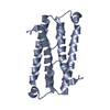 1rfyS S: Starting model for refinement |
|---|---|
| Similar structure data |


-Links
 PDBj
PDBj- Assembly
| Deposited unit | 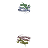
| ||||||||
|---|---|---|---|---|---|---|---|---|---|
| 1 | 
| ||||||||
| 2 | 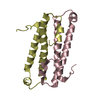
| ||||||||
| Unit cell |
| ||||||||
| Details | The biological assembly is a homodimer, consisting of either chains A and B, or chains C and D |
-Components
| #1: Protein | Mass: 11141.806 Da / Num. of mol.: 4 Source method: isolated from a genetically manipulated source Source: (gene. exp.) Agrobacterium tumefaciens (bacteria) / Gene: traM2 / Plasmid: pET15b / Species (production host): Escherichia coli / Production host: #2: Water | ChemComp-HOH / |  Mass: 18.015 Da / Num. of mol.: 307 / Source method: isolated from a natural source / Formula: H2O Mass: 18.015 Da / Num. of mol.: 307 / Source method: isolated from a natural source / Formula: H2O |
|---|
-Experimental details
-Experiment
| Experiment | Method: X-RAY DIFFRACTION / Number of used crystals: 1 |
|---|
- Sample preparation
| Crystal | Density Matthews: 3.2 Å3/Da / Density % sol: 61.56 % |
|---|---|
| Crystal grow | Temperature: 298 K / Method: vapor diffusion, hanging drop / pH: 8 Details: 50 mM TrisHCl, 200 mM NaCl, 0.5 mM EDTA, 1 mM DTT, 25% ethylene glycol, pH 8, VAPOR DIFFUSION, HANGING DROP, temperature 298K |
-Data collection
| Diffraction | Mean temperature: 298 K |
|---|---|
| Diffraction source | Source: SYNCHROTRON / Site: ALS  / Beamline: 5.0.2 / Beamline: 5.0.2 |
| Detector | Type: ADSC QUANTUM 4 / Detector: CCD / Date: Oct 31, 2004 |
| Radiation | Monochromator: Si 111 Channel / Protocol: SINGLE WAVELENGTH / Monochromatic (M) / Laue (L): M / Scattering type: x-ray |
| Radiation wavelength | Relative weight: 1 |
| Reflection | Resolution: 2.1→40 Å / Num. all: 32820 / Num. obs: 32632 / % possible obs: 99.4 % / Observed criterion σ(F): 1 / Observed criterion σ(I): 1 |
| Reflection shell | Resolution: 2.1→2.18 Å / % possible all: 99.7 |
- Processing
| Software |
| ||||||||||||||||||||
|---|---|---|---|---|---|---|---|---|---|---|---|---|---|---|---|---|---|---|---|---|---|
| Refinement | Method to determine structure: MOLECULAR REPLACEMENT Starting model: PDB entry 1RFY Resolution: 2.1→40 Å / Cross valid method: THROUGHOUT / σ(F): 0 / Stereochemistry target values: Engh & Huber
| ||||||||||||||||||||
| Refine analyze |
| ||||||||||||||||||||
| Refinement step | Cycle: LAST / Resolution: 2.1→40 Å
| ||||||||||||||||||||
| Refine LS restraints |
| ||||||||||||||||||||
| LS refinement shell | Resolution: 2.1→2.23 Å / Rfactor Rfree error: 0.016
|
