 Movie
Movie Controller

[English] 日本語
 Yorodumi
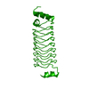
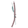
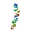
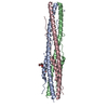
- PDB-4nkj: Structure of influenza B virus hemagglutinin at membrane fusion pH -
+ Open data

- Basic information
| Entry | Database: PDB / ID: 4nkj | ||||||
|---|---|---|---|---|---|---|---|
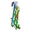
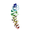
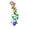
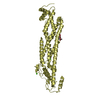
| Title | Structure of influenza B virus hemagglutinin at membrane fusion pH | ||||||
 Components Components | Hemagglutinin HA2 | ||||||
 Keywords Keywords | VIRAL PROTEIN / HA | ||||||
| Function / homology |  Function and homology information Function and homology informationviral budding from plasma membrane / host cell surface receptor binding / endocytosis involved in viral entry into host cell / fusion of virus membrane with host plasma membrane / fusion of virus membrane with host endosome membrane / viral envelope / virion attachment to host cell / host cell plasma membrane / virion membrane / membrane Similarity search - Function | ||||||
| Biological species |  Influenza B virus Influenza B virus | ||||||
| Method |  X-RAY DIFFRACTION / SYNCHROTRON / MOLECULAR REPLACEMENT / Resolution: 2.4535 Å X-RAY DIFFRACTION / SYNCHROTRON / MOLECULAR REPLACEMENT / Resolution: 2.4535 Å | ||||||
 Authors Authors | Ni, F. / Chen, X. / Shen, J. / Wang, Q. | ||||||
 Citation Citation | Journal: Biochemistry / Year: 2014 Title: Structural insights into the membrane fusion mechanism mediated by influenza virus hemagglutinin. Authors: Ni, F. / Chen, X. / Shen, J. / Wang, Q. | ||||||
| History |
|
- Structure visualization
| Structure viewer | Molecule: MolmilJmol/JSmol |
|---|
- Downloads & links
-Download
| PDBx/mmCIF format | 4nkj.cif.gz | 38.7 KB | Display | PDBx/mmCIF format |
|---|---|---|---|---|
| PDB format | pdb4nkj.ent.gz | 27.3 KB | Display | PDB format |
| PDBx/mmJSON format | 4nkj.json.gz | Tree view | PDBx/mmJSON format | |
| Others |  Other downloads Other downloads |
-Validation report
| Arichive directory | https://data.pdbj.org/pub/pdb/validation_reports/nk/4nkjftp://data.pdbj.org/pub/pdb/validation_reports/nk/4nkj | HTTPS FTP |
|---|
-Related structure data
| Similar structure data |
|---|


-Links
 PDBj
PDBj


- Assembly
| Deposited unit | 
| ||||||||
|---|---|---|---|---|---|---|---|---|---|
| 1 | 
| ||||||||
| Unit cell |
| ||||||||
| Components on special symmetry positions |
|
-Components
| #1: Protein | Mass: 17211.225 Da / Num. of mol.: 1 / Fragment: Hemagglutinin HA2 (UNP Residues 392-541) Source method: isolated from a genetically manipulated source Source: (gene. exp.) Influenza B virus / Gene: HA / Production host:  |
|---|---|
| #2: Water | ChemComp-HOH /  Mass: 18.015 Da / Num. of mol.: 27 / Source method: isolated from a natural source / Formula: H2O Mass: 18.015 Da / Num. of mol.: 27 / Source method: isolated from a natural source / Formula: H2O |
| Has protein modification | Y |
-Experimental details
-Experiment
| Experiment | Method: X-RAY DIFFRACTION / Number of used crystals: 1 |
|---|
- Sample preparation
| Crystal | Density Matthews: 2.3 Å3/Da / Density % sol: 46.59 % |
|---|
-Data collection
| Diffraction | Mean temperature: 200 K |
|---|---|
| Diffraction source | Source: SYNCHROTRON / Site: APS  / Beamline: 21-ID-D / Wavelength: 1.127 Å / Beamline: 21-ID-D / Wavelength: 1.127 Å |
| Detector | Type: MARMOSAIC 225 mm CCD / Detector: CCD |
| Radiation | Protocol: SINGLE WAVELENGTH / Monochromatic (M) / Laue (L): M / Scattering type: x-ray |
| Radiation wavelength | Wavelength: 1.127 Å / Relative weight: 1 |
| Reflection | Resolution: 2.45→37.76 Å / Num. all: 6281 / Num. obs: 5617 / % possible obs: 89.4 % / Observed criterion σ(F): 2 / Observed criterion σ(I): 1 |
| Reflection shell | Resolution: 2.45→2.54 Å / Redundancy: 4.4 % / Rmerge(I) obs: 0.21 / Mean I/σ(I) obs: 5.21 / Num. unique all: 615 / % possible all: 94.96 |
- Processing
| Software |
| ||||||||||||||||||||||||
|---|---|---|---|---|---|---|---|---|---|---|---|---|---|---|---|---|---|---|---|---|---|---|---|---|---|
| Refinement | Method to determine structure: MOLECULAR REPLACEMENT / Resolution: 2.4535→37.758 Å / SU ML: 0.27 / σ(F): 1.94 / Phase error: 32.86 / Stereochemistry target values: ML
| ||||||||||||||||||||||||
| Solvent computation | Shrinkage radii: 0.61 Å / VDW probe radii: 0.9 Å / Solvent model: FLAT BULK SOLVENT MODEL / Bsol: 60.41 Å2 / ksol: 0.358 e/Å3 | ||||||||||||||||||||||||
| Displacement parameters |
| ||||||||||||||||||||||||
| Refinement step | Cycle: LAST / Resolution: 2.4535→37.758 Å
| ||||||||||||||||||||||||
| Refine LS restraints |
| ||||||||||||||||||||||||
| LS refinement shell |
|
