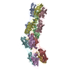
[English] 日本語
Yorodumi
- PDB-5cyl: Crystal structure of the CupB6 tip adhesin from Pseudomonas aeruginosa -

+
Open data


ID or keywords:

Loading...

-
Basic information

Entry
Database: PDB / ID: 5cyl
TitleCrystal structure of the CupB6 tip adhesin from Pseudomonas aeruginosa
ComponentsFimbrial subunit CupB6
KeywordsCELL ADHESION / biofilm / adhesion / chaperone-usher / pilin
Function / homologypilus organization / cell adhesion involved in single-species biofilm formation / Fimbrial-type adhesion domain / Fimbrial protein / Fimbrial-type adhesion domain superfamily / Adhesion domain superfamily / pilus / Fimbrial subunit CupB6
Function and homology information
Biological speciesPseudomonas aeruginosa (bacteria)
MethodX-RAY DIFFRACTION / SYNCHROTRON / MAD / Resolution: 2.77 Å
AuthorsRasheed, M. / Garnett, J.A. / Perez-Dorado, I. / Matthews, S.J.
Funding support United Kingdom, 1items
OrganizationGrant numberCountry
Wellcome Trust100280 United Kingdom
CitationJournal: Biochim.Biophys.Acta / Year: 2016
Title: Crystal structure of the CupB6 adhesive tip from the chaperone-usher family of pili from Pseudomonas aeruginosa.
Authors: Rasheed, M. / Garnett, J. / Perez-Dorado, I. / Muhl, D. / Filloux, A. / Matthews, S.
History
DepositionJul 30, 2015Deposition site: PDBE / Processing site: PDBE
Revision 1.0Oct 5, 2016Provider: repository / Type: Initial release

-
Structure visualization

Structure viewerMolecule:
MolmilJmol/JSmol

Downloads & links

-
Assembly

Deposited unit
A: Fimbrial subunit CupB6
B: Fimbrial subunit CupB6
C: Fimbrial subunit CupB6
D: Fimbrial subunit CupB6
E: Fimbrial subunit CupB6
F: Fimbrial subunit CupB6
G: Fimbrial subunit CupB6
H: Fimbrial subunit CupB6


Theoretical massNumber of molelcules
Total (without water)313,7618
Polymers313,7618
Non-polymers00
Water5,368298
1
A: Fimbrial subunit CupB6


Theoretical massNumber of molelcules
Total (without water)39,2201
Polymers39,2201
Non-polymers00
Water181
TypeNameSymmetry operationNumber
identity operation1_555x,y,z1
2
B: Fimbrial subunit CupB6


Theoretical massNumber of molelcules
Total (without water)39,2201
Polymers39,2201
Non-polymers00
Water181
TypeNameSymmetry operationNumber
identity operation1_555x,y,z1
3
C: Fimbrial subunit CupB6


Theoretical massNumber of molelcules
Total (without water)39,2201
Polymers39,2201
Non-polymers00
Water181
TypeNameSymmetry operationNumber
identity operation1_555x,y,z1
4
D: Fimbrial subunit CupB6


Theoretical massNumber of molelcules
Total (without water)39,2201
Polymers39,2201
Non-polymers00
Water181
TypeNameSymmetry operationNumber
identity operation1_555x,y,z1
5
E: Fimbrial subunit CupB6


Theoretical massNumber of molelcules
Total (without water)39,2201
Polymers39,2201
Non-polymers00
Water181
TypeNameSymmetry operationNumber
identity operation1_555x,y,z1
6
F: Fimbrial subunit CupB6


Theoretical massNumber of molelcules
Total (without water)39,2201
Polymers39,2201
Non-polymers00
Water181
TypeNameSymmetry operationNumber
identity operation1_555x,y,z1
7
G: Fimbrial subunit CupB6


Theoretical massNumber of molelcules
Total (without water)39,2201
Polymers39,2201
Non-polymers00
Water181
TypeNameSymmetry operationNumber
identity operation1_555x,y,z1
8
H: Fimbrial subunit CupB6


Theoretical massNumber of molelcules
Total (without water)39,2201
Polymers39,2201
Non-polymers00
Water181
TypeNameSymmetry operationNumber
identity operation1_555x,y,z1
Unit cell
Length a, b, c (Å)358.813, 88.930, 172.974
Angle α, β, γ (deg.)90.00, 112.94, 90.00
Int Tables number5
Space group name H-MC121
Noncrystallographic symmetry (NCS)NCS domain:
IDEns-IDDetails
11A
21B
12A
22C
13A
23D
14A
24E
15A
25F
16A
26G
17A
27H
18B
28C
19B
29D
110B
210E
111B
211F
112B
212G
113B
213H
114C
214D
115C
215E
116C
216F
117C
217G
118C
218H
119D
219E
120D
220F
121D
221G
122D
222H
123E
223F
124E
224G
125E
225H
126F
226G
127F
227H
128G
228H

NCS domain segments:
Dom-IDComponent-IDEns-IDRefine codeAuth asym-IDAuth seq-ID
1010A3 - 361
2010B3 - 361
1020A4 - 360
2020C4 - 360
1030A3 - 360
2030D3 - 360
1040A4 - 363
2040E4 - 363
1050A3 - 361
2050F3 - 361
1060A3 - 360
2060G3 - 360
1070A3 - 363
2070H3 - 363
1080B4 - 360
2080C4 - 360
1090B1 - 361
2090D1 - 361
10100B4 - 360
20100E4 - 360
10110B3 - 361
20110F3 - 361
10120B1 - 360
20120G1 - 360
10130B1 - 361
20130H1 - 361
10140C4 - 360
20140D4 - 360
10150C4 - 360
20150E4 - 360
10160C4 - 360
20160F4 - 360
10170C4 - 360
20170G4 - 360
10180C4 - 360
20180H4 - 360
10190D4 - 361
20190E4 - 361
10200D3 - 361
20200F3 - 361
10210D1 - 360
20210G1 - 360
10220D1 - 361
20220H1 - 361
10230E4 - 361
20230F4 - 361
10240E4 - 359
20240G4 - 359
10250E4 - 362
20250H4 - 362
10260F3 - 360
20260G3 - 360
10270F3 - 361
20270H3 - 361
10280G1 - 360
20280H1 - 360

NCS ensembles :
ID
1
2
3
4
5
6
7
8
9
10
11
12
13
14
15
16
17
18
19
20
21
22
23
24
25
26
27
28

-
Components

#1: Protein
Fimbrial subunit CupB6


Mass: 39220.172 Da / Num. of mol.: 8 / Fragment: UNP residues 38-381
Source method: isolated from a genetically manipulated source
Source: (gene. exp.) Pseudomonas aeruginosa (bacteria) / Gene: cupB6, PA4081 / Production host: Escherichia coli (E. coli) / References: UniProt: Q9HWU7
#2: Water ChemComp-HOH / water / Water


Mass: 18.015 Da / Num. of mol.: 298 / Source method: isolated from a natural source / Formula: H2O

-
Experimental details

-
Experiment

ExperimentMethod: X-RAY DIFFRACTION / Number of used crystals: 1

-
Sample preparation

CrystalDensity Matthews: 4.07 Å3/Da / Density % sol: 69.79 %
Crystal growTemperature: 291.15 K / Method: vapor diffusion, hanging drop / pH: 4.2
Details: 1.6M NaH2PO4, 400 mM KH2PO4, 100mM phosphate citrate

-
Data collection

DiffractionMean temperature: 100 K
Diffraction sourceSource: SYNCHROTRON / Site: Diamond / Beamline: I02 / Wavelength: 0.92002, 0.97903, 0.97957, 0.96757
DetectorType: DECTRIS PILATUS 6M / Detector: PIXEL / Date: Jan 19, 2014
RadiationProtocol: SINGLE WAVELENGTH / Monochromatic (M) / Laue (L): M / Scattering type: x-ray
Radiation wavelength
IDWavelength (Å)Relative weight
10.920021
20.979031
30.979571
40.967571
ReflectionResolution: 2.77→97.29 Å / Num. obs: 416327 / % possible obs: 96.6 % / Redundancy: 3.4 % / Biso Wilson estimate: 57.4 Å2 / CC1/2: 0.97 / Rmerge(I) obs: 0.098 / Net I/σ(I): 8.9
Reflection shellResolution: 2.77→2.84 Å / Redundancy: 3.3 % / Rmerge(I) obs: 0.857 / Mean I/σ(I) obs: 1.4 / CC1/2: 0.578 / % possible all: 96.9

-
Processing

Software
NameVersionClassification
REFMAC5.8.0103refinement
SCALAdata scaling
XDSdata reduction
SHARPphasing
RefinementMethod to determine structure: MAD / Resolution: 2.77→97.29 Å / Cor.coef. Fo:Fc: 0.916 / Cor.coef. Fo:Fc free: 0.898 / Cross valid method: THROUGHOUT / ESU R: 0.541 / ESU R Free: 0.322 / Stereochemistry target values: MAXIMUM LIKELIHOOD / Details: HYDROGENS HAVE BEEN ADDED IN THE RIDING POSITIONS
RfactorNum. reflection% reflectionSelection details
Rfree0.26645 6115 5 %RANDOM
Rwork0.23844 ---
obs0.23983 117132 96.17 %-
Solvent computationIon probe radii: 0.8 Å / Shrinkage radii: 0.8 Å / VDW probe radii: 1.2 Å / Solvent model: MASK
Displacement parametersBiso mean: 55.613 Å2
Baniso -1Baniso -2Baniso -3
1--0.26 Å20 Å2-0.04 Å2
2--0.16 Å2-0 Å2
3---0.1 Å2
Refinement stepCycle: LAST / Resolution: 2.77→97.29 Å
ProteinNucleic acidLigandSolventTotal
Num. atoms20683 0 0 298 20981
Refine LS restraints
Refine-IDTypeDev idealDev ideal targetNumber
X-RAY DIFFRACTIONr_bond_refined_d0.0130.01921100
X-RAY DIFFRACTIONr_bond_other_d00.0219922
X-RAY DIFFRACTIONr_angle_refined_deg1.7461.97428772
X-RAY DIFFRACTIONr_angle_other_deg3.744345856
X-RAY DIFFRACTIONr_dihedral_angle_1_deg5.13552810
X-RAY DIFFRACTIONr_dihedral_angle_2_deg35.624.277788
X-RAY DIFFRACTIONr_dihedral_angle_3_deg18.675153105
X-RAY DIFFRACTIONr_dihedral_angle_4_deg18.56715116
X-RAY DIFFRACTIONr_chiral_restr0.0870.23321
X-RAY DIFFRACTIONr_gen_planes_refined0.0070.02124248
X-RAY DIFFRACTIONr_gen_planes_other0.0080.024456
X-RAY DIFFRACTIONr_nbd_refined
X-RAY DIFFRACTIONr_nbd_other
X-RAY DIFFRACTIONr_nbtor_refined
X-RAY DIFFRACTIONr_nbtor_other
X-RAY DIFFRACTIONr_xyhbond_nbd_refined
X-RAY DIFFRACTIONr_xyhbond_nbd_other
X-RAY DIFFRACTIONr_metal_ion_refined
X-RAY DIFFRACTIONr_metal_ion_other
X-RAY DIFFRACTIONr_symmetry_vdw_refined
X-RAY DIFFRACTIONr_symmetry_vdw_other
X-RAY DIFFRACTIONr_symmetry_hbond_refined
X-RAY DIFFRACTIONr_symmetry_hbond_other
X-RAY DIFFRACTIONr_symmetry_metal_ion_refined
X-RAY DIFFRACTIONr_symmetry_metal_ion_other
X-RAY DIFFRACTIONr_mcbond_it4.2785.73911320
X-RAY DIFFRACTIONr_mcbond_other4.2785.73911319
X-RAY DIFFRACTIONr_mcangle_it6.7578.59414102
X-RAY DIFFRACTIONr_mcangle_other6.7578.59414103
X-RAY DIFFRACTIONr_scbond_it4.6555.7269780
X-RAY DIFFRACTIONr_scbond_other4.6525.7259778
X-RAY DIFFRACTIONr_scangle_it
X-RAY DIFFRACTIONr_scangle_other7.1128.50114670
X-RAY DIFFRACTIONr_long_range_B_refined10.97445.35922283
X-RAY DIFFRACTIONr_long_range_B_other10.97345.33922256
X-RAY DIFFRACTIONr_rigid_bond_restr
X-RAY DIFFRACTIONr_sphericity_free
X-RAY DIFFRACTIONr_sphericity_bonded
Refine LS restraints NCS

Refine-ID: X-RAY DIFFRACTION / Type: interatomic distance / Weight position: 0.05

Ens-IDDom-IDAuth asym-IDNumberRms dev position (Å)
11A348160.23
12B348160.23
21A340700.23
22C340700.23
31A336580.24
32D336580.24
41A352540.22
42E352540.22
51A309020.25
52F309020.25
61A326160.24
62G326160.24
71A356560.22
72H356560.22
81B331080.24
82C331080.24
91B339640.24
92D339640.24
101B343840.23
102E343840.23
111B318360.24
112F318360.24
121B332760.25
122G332760.25
131B357500.23
132H357500.23
141C329920.25
142D329920.25
151C338880.23
152E338880.23
161C303800.24
162F303800.24
171C320020.25
172G320020.25
181C338940.23
182H338940.23
191D333080.24
192E333080.24
201D317220.24
202F317220.24
211D326180.25
212G326180.25
221D341020.24
222H341020.24
231E314740.24
232F314740.24
241E326120.24
242G326120.24
251E354680.22
252H354680.22
261F308300.24
262G308300.24
271F321200.24
272H321200.24
281G331340.25
282H331340.25
LS refinement shellResolution: 2.77→2.842 Å / Total num. of bins used: 20
RfactorNum. reflection% reflection
Rfree0.426 450 -
Rwork0.397 8605 -
obs--96.35 %

+
About Yorodumi

-
News

-
Feb 9, 2022. New format data for meta-information of EMDB entries

New format data for meta-information of EMDB entries

  • Version 3 of the EMDB header file is now the official format.
  • The previous official version 1.9 will be removed from the archive.

Related info.:EMDB header

External links:wwPDB to switch to version 3 of the EMDB data model

-
Aug 12, 2020. Covid-19 info

Covid-19 info

URL: https://pdbj.org/emnavi/covid19.php

New page: Covid-19 featured information page in EM Navigator.

Related info.:Covid-19 info / Mar 5, 2020. Novel coronavirus structure data

+
Mar 5, 2020. Novel coronavirus structure data

Novel coronavirus structure data

Related info.:Yorodumi Speices / Aug 12, 2020. Covid-19 info

External links:COVID-19 featured content - PDBj / Molecule of the Month (242):Coronavirus Proteases

+
Jan 31, 2019. EMDB accession codes are about to change! (news from PDBe EMDB page)

EMDB accession codes are about to change! (news from PDBe EMDB page)

  • The allocation of 4 digits for EMDB accession codes will soon come to an end. Whilst these codes will remain in use, new EMDB accession codes will include an additional digit and will expand incrementally as the available range of codes is exhausted. The current 4-digit format prefixed with “EMD-” (i.e. EMD-XXXX) will advance to a 5-digit format (i.e. EMD-XXXXX), and so on. It is currently estimated that the 4-digit codes will be depleted around Spring 2019, at which point the 5-digit format will come into force.
  • The EM Navigator/Yorodumi systems omit the EMD- prefix.

Related info.:Q: What is EMD? / ID/Accession-code notation in Yorodumi/EM Navigator

External links:EMDB Accession Codes are Changing Soon! / Contact to PDBj

+
Jul 12, 2017. Major update of PDB

Major update of PDB

  • wwPDB released updated PDB data conforming to the new PDBx/mmCIF dictionary.
  • This is a major update changing the version number from 4 to 5, and with Remediation, in which all the entries are updated.
  • In this update, many items about electron microscopy experimental information are reorganized (e.g. em_software).
  • Now, EM Navigator and Yorodumi are based on the updated data.

External links:wwPDB Remediation / Enriched Model Files Conforming to OneDep Data Standards Now Available in the PDB FTP Archive

-
Yorodumi

Thousand views of thousand structures

  • Yorodumi is a browser for structure data from EMDB, PDB, SASBDB, etc.
  • This page is also the successor to EM Navigator detail page, and also detail information page/front-end page for Omokage search.
  • The word "yorodu" (or yorozu) is an old Japanese word meaning "ten thousand". "mi" (miru) is to see.

Related info.:EMDB / PDB / SASBDB / Comparison of 3 databanks / Yorodumi Search / Aug 31, 2016. New EM Navigator & Yorodumi / Yorodumi Papers / Jmol/JSmol / Function and homology information / Changes in new EM Navigator and Yorodumi

Read more