 Movie
Movie Controller

[English] 日本語
 Yorodumi
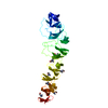
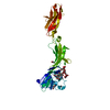
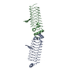
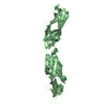
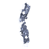
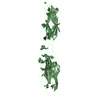
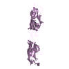
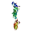
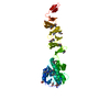
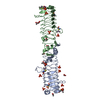
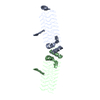
- PDB-2v05: CRYSTAL STRUCTURE OF CHOLINE BINDING PROTEIN F FROM STREPTOCOCCUS... -
+ Open data

- Basic information
| Entry | Database: PDB / ID: 2v05 | ||||||
|---|---|---|---|---|---|---|---|
| Title | CRYSTAL STRUCTURE OF CHOLINE BINDING PROTEIN F FROM STREPTOCOCCUS PNEUMONIAE. CRYSTAL FORM II. | ||||||
 Components Components | CHOLINE BINDING PROTEIN F | ||||||
 Keywords Keywords | LIPID BINDING PROTEIN / CBPF / CHOLINE-BINDING-PROTEIN / LIPID-BINDING-PROTEIN | ||||||
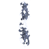
| Function / homology |  Function and homology information Function and homology informationleft handed beta-beta-3-solenoid - #20 / Cholin Binding / left handed beta-beta-3-solenoid / Choline-binding repeat / Putative cell wall binding repeat / Cell wall/choline-binding repeat / Cell wall-binding repeat profile. / Ribbon / Mainly Beta Similarity search - Domain/homology | ||||||
| Biological species |   STREPTOCOCCUS PNEUMONIAE (bacteria) STREPTOCOCCUS PNEUMONIAE (bacteria) | ||||||
| Method |  X-RAY DIFFRACTION / SAD / Resolution: 1.67 Å X-RAY DIFFRACTION / SAD / Resolution: 1.67 Å | ||||||
 Authors Authors | Hermoso, J. / Molina, R. / Kahn, R. / Stelter, M. | ||||||
 Citation Citation | Journal: To be Published Title: Crystal Structure of the Choline Binding Protein F from Streptococcus Pneumoniae Authors: Hermoso, J. / Molina, R. / Gonzalez, A. / Stelter, M. / Garcia, P. / Kahn, R. / Martinez-Ripoll, M. / Garcia, J.L. / Menendez, M. | ||||||
| History |
|
- Structure visualization
| Structure viewer | Molecule: MolmilJmol/JSmol |
|---|
- Downloads & links
-Download
| PDBx/mmCIF format | 2v05.cif.gz | 85.6 KB | Display | PDBx/mmCIF format |
|---|---|---|---|---|
| PDB format | pdb2v05.ent.gz | 65.5 KB | Display | PDB format |
| PDBx/mmJSON format | 2v05.json.gz | Tree view | PDBx/mmJSON format | |
| Others |  Other downloads Other downloads |
-Validation report
| Arichive directory | https://data.pdbj.org/pub/pdb/validation_reports/v0/2v05ftp://data.pdbj.org/pub/pdb/validation_reports/v0/2v05 | HTTPS FTP |
|---|
-Related structure data
| Related structure data | |
|---|---|
| Similar structure data |
-Links
 PDBj
PDBj
- Assembly
| Deposited unit | 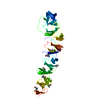
| ||||||||
|---|---|---|---|---|---|---|---|---|---|
| 1 |
| ||||||||
| Unit cell |
|
-Components
| #1: Protein | Mass: 36322.938 Da / Num. of mol.: 1 Source method: isolated from a genetically manipulated source Source: (gene. exp.) STREPTOCOCCUS PNEUMONIAE (bacteria) / Strain: R6 / Production host: | ||
|---|---|---|---|
| #2: Chemical | ChemComp-CHT / 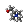  Mass: 104.171 Da / Num. of mol.: 7 / Source method: obtained synthetically / Formula: C5H14NO Mass: 104.171 Da / Num. of mol.: 7 / Source method: obtained synthetically / Formula: C5H14NO#3: Water | ChemComp-HOH / |  Mass: 18.015 Da / Num. of mol.: 490 / Source method: isolated from a natural source / Formula: H2O Mass: 18.015 Da / Num. of mol.: 490 / Source method: isolated from a natural source / Formula: H2O |
-Experimental details
-Experiment
| Experiment | Method: X-RAY DIFFRACTION / Number of used crystals: 1 |
|---|
- Sample preparation
| Crystal | Density Matthews: 2.94 Å3/Da / Density % sol: 58 % / Description: NONE |
|---|---|
| Crystal grow | pH: 8.5 / Details: pH 8.5 |
-Data collection
| Diffraction | Mean temperature: 291 K |
|---|---|
| Diffraction source | Source: ROTATING ANODE / Type: ENRAF-NONIUS / Wavelength: 1.5418 |
| Detector | Type: MARRESEARCH / Detector: IMAGE PLATE / Date: Feb 6, 2004 / Details: DOUBLE-MIRROR |
| Radiation | Monochromator: GRAPHITE / Protocol: SINGLE WAVELENGTH / Monochromatic (M) / Laue (L): M / Scattering type: x-ray |
| Radiation wavelength | Wavelength: 1.5418 Å / Relative weight: 1 |
| Reflection | Resolution: 1.67→21.07 Å / Num. obs: 50379 / % possible obs: 99.1 % / Observed criterion σ(I): 2 / Redundancy: 12.4 % / Rmerge(I) obs: 0.05 / Net I/σ(I): 9.3 |
| Reflection shell | Resolution: 1.67→1.76 Å / Redundancy: 11.5 % / Rmerge(I) obs: 0.34 / Mean I/σ(I) obs: 2.2 / % possible all: 96.7 |
- Processing
| Software |
| ||||||||||||||||||||||||||||||||||||||||||||||||||||||||||||||||||||||||||||||||||||||||||||||||||||||||||||||||||||||||||||||||||||||||||||||||||||||||||||||||||||||||||||||||||||||
|---|---|---|---|---|---|---|---|---|---|---|---|---|---|---|---|---|---|---|---|---|---|---|---|---|---|---|---|---|---|---|---|---|---|---|---|---|---|---|---|---|---|---|---|---|---|---|---|---|---|---|---|---|---|---|---|---|---|---|---|---|---|---|---|---|---|---|---|---|---|---|---|---|---|---|---|---|---|---|---|---|---|---|---|---|---|---|---|---|---|---|---|---|---|---|---|---|---|---|---|---|---|---|---|---|---|---|---|---|---|---|---|---|---|---|---|---|---|---|---|---|---|---|---|---|---|---|---|---|---|---|---|---|---|---|---|---|---|---|---|---|---|---|---|---|---|---|---|---|---|---|---|---|---|---|---|---|---|---|---|---|---|---|---|---|---|---|---|---|---|---|---|---|---|---|---|---|---|---|---|---|---|---|---|
| Refinement | Method to determine structure: SAD Starting model: NONE Resolution: 1.67→76.7 Å / Cor.coef. Fo:Fc: 0.95 / Cor.coef. Fo:Fc free: 0.94 / SU B: 1.766 / SU ML: 0.061 / TLS residual ADP flag: LIKELY RESIDUAL / Cross valid method: THROUGHOUT / ESU R: 0.101 / ESU R Free: 0.098 / Stereochemistry target values: MAXIMUM LIKELIHOOD Details: HYDROGENS HAVE BEEN ADDED IN THE RIDING POSITIONS. RESIDUES FROM 147 TO 150 AND FROM 169 TO 177, ARE DISORDERED. THEY DO NOT APPEAR IN THE MODEL DISORDERED REGIONS FROM RESIDUES 147 TO 150 ...Details: HYDROGENS HAVE BEEN ADDED IN THE RIDING POSITIONS. RESIDUES FROM 147 TO 150 AND FROM 169 TO 177, ARE DISORDERED. THEY DO NOT APPEAR IN THE MODEL DISORDERED REGIONS FROM RESIDUES 147 TO 150 AND FROM 169 TO 177. THEY DO NOT APPEAR IN THE MODEL.
| ||||||||||||||||||||||||||||||||||||||||||||||||||||||||||||||||||||||||||||||||||||||||||||||||||||||||||||||||||||||||||||||||||||||||||||||||||||||||||||||||||||||||||||||||||||||
| Solvent computation | Ion probe radii: 0.8 Å / Shrinkage radii: 0.8 Å / VDW probe radii: 1.4 Å / Solvent model: BABINET MODEL WITH MASK | ||||||||||||||||||||||||||||||||||||||||||||||||||||||||||||||||||||||||||||||||||||||||||||||||||||||||||||||||||||||||||||||||||||||||||||||||||||||||||||||||||||||||||||||||||||||
| Displacement parameters | Biso mean: 20.58 Å2
| ||||||||||||||||||||||||||||||||||||||||||||||||||||||||||||||||||||||||||||||||||||||||||||||||||||||||||||||||||||||||||||||||||||||||||||||||||||||||||||||||||||||||||||||||||||||
| Refinement step | Cycle: LAST / Resolution: 1.67→76.7 Å
| ||||||||||||||||||||||||||||||||||||||||||||||||||||||||||||||||||||||||||||||||||||||||||||||||||||||||||||||||||||||||||||||||||||||||||||||||||||||||||||||||||||||||||||||||||||||
| Refine LS restraints |
|