| Entry | Database: PDB / ID: 2v04
|
|---|
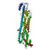
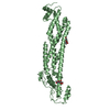
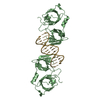
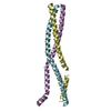
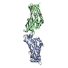
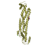
| Title | CRYSTAL STRUCTURE OF CHOLINE BINDING PROTEIN F FROM STREPTOCOCCUS PNEUMONIAE |
|---|
 Components Components | CHOLINE BINDING PROTEIN F |
|---|
 Keywords Keywords | LIPID BINDING PROTEIN / CBPF / CHOLINE-BINDING-PROTEIN / LIPID-BINDING-PROTEIN |
|---|
| Function / homology |  Function and homology information Function and homology information
left handed beta-beta-3-solenoid - #20 / Cholin Binding / left handed beta-beta-3-solenoid / Choline-binding repeat / Putative cell wall binding repeat / Cell wall/choline-binding repeat / Cell wall-binding repeat profile. / Ribbon / Mainly BetaSimilarity search - Domain/homology |
|---|
| Biological species |   STREPTOCOCCUS PNEUMONIAE (bacteria) STREPTOCOCCUS PNEUMONIAE (bacteria) |
|---|
| Method |  X-RAY DIFFRACTION / SYNCHROTRON / SAD / Resolution: 2.1 Å X-RAY DIFFRACTION / SYNCHROTRON / SAD / Resolution: 2.1 Å |
|---|
 Authors Authors | Hermoso, J. / Molina, R. / Kahn, R. / Stelter, M. |
|---|
 Citation Citation | Journal: Embo Rep. / Year: 2009
Title: Crystal Structure of Cbpf, a Bifunctional Choline-Binding Protein and Autolysis Regulator from Streptococcus Pneumoniae.
Authors: Molina, R. / Gonzalez, A. / Stelter, M. / Perez-Dorado, I. / Kahn, R. / Morales, M. / Campuzano, S. / Campillo, N.E. / Mobashery, S. / Garcia, J.L. / Garcia, P. / Hermoso, J.A. |
|---|
| History | | Deposition | May 8, 2007 | Deposition site: PDBE / Processing site: PDBE |
|---|
| Revision 1.0 | Jun 10, 2008 | Provider: repository / Type: Initial release |
|---|
| Revision 1.1 | Jul 13, 2011 | Group: Advisory / Refinement description / Version format compliance |
|---|
| Revision 1.2 | Apr 25, 2018 | Group: Data collection / Database references / Category: citation / diffrn_source
Item: _citation.title / _diffrn_source.pdbx_synchrotron_beamline ..._citation.title / _diffrn_source.pdbx_synchrotron_beamline / _diffrn_source.pdbx_synchrotron_site / _diffrn_source.source / _diffrn_source.type |
|---|
| Revision 2.0 | May 8, 2024 | Group: Atomic model / Data collection ...Atomic model / Data collection / Database references / Derived calculations / Other
Category: atom_site / chem_comp_atom ...atom_site / chem_comp_atom / chem_comp_bond / database_2 / pdbx_database_status / struct_site
Item: _atom_site.occupancy / _database_2.pdbx_DOI ..._atom_site.occupancy / _database_2.pdbx_DOI / _database_2.pdbx_database_accession / _pdbx_database_status.status_code_sf / _struct_site.pdbx_auth_asym_id / _struct_site.pdbx_auth_comp_id / _struct_site.pdbx_auth_seq_id |
|---|
|
|---|
 Movie
Movie Controller

 Yorodumi
 Open data
 Basic information
 Structure visualization
 Downloads & links
 Other downloads

 PDBj
PDBj
 Assembly
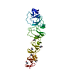
 Mass: 104.171 Da / Num. of mol.: 7 / Source method: obtained synthetically / Formula: C5H14NO
 Mass: 18.015 Da / Num. of mol.: 310 / Source method: isolated from a natural source / Formula: H2O
 Sample preparation
 / Beamline: ID29 / Wavelength: 1.5418
  Processing