 Movie
Movie Controller

[English] 日本語
 Yorodumi
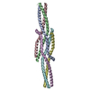
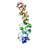
- PDB-6gbp: Crystal Structure of the oligomerization domain of VP35 from Ebol... -
+ Open data

- Basic information
| Entry | Database: PDB / ID: 6gbp | ||||||
|---|---|---|---|---|---|---|---|
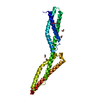
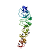
| Title | Crystal Structure of the oligomerization domain of VP35 from Ebola virus, mercury derivative | ||||||
 Components Components | Polymerase cofactor VP35 | ||||||
 Keywords Keywords | VIRAL PROTEIN / coiled-coil | ||||||
| Function / homology |  Function and homology information Function and homology informationsymbiont-mediated suppression of host immune response / symbiont-mediated suppression of host RNAi-mediated antiviral immune response / negative regulation of miRNA-mediated gene silencing / symbiont-mediated suppression of host cytoplasmic pattern recognition receptor signaling pathway via inhibition of IKBKE activity / symbiont-mediated suppression of host cytoplasmic pattern recognition receptor signaling pathway via inhibition of IRF7 activity / symbiont-mediated suppression of host antigen processing and presentation of peptide antigen via MHC class II / symbiont-mediated suppression of host PKR/eIFalpha signaling / positive regulation of protein sumoylation / viral transcription / molecular sequestering activity ...symbiont-mediated suppression of host immune response / symbiont-mediated suppression of host RNAi-mediated antiviral immune response / negative regulation of miRNA-mediated gene silencing / symbiont-mediated suppression of host cytoplasmic pattern recognition receptor signaling pathway via inhibition of IKBKE activity / symbiont-mediated suppression of host cytoplasmic pattern recognition receptor signaling pathway via inhibition of IRF7 activity / symbiont-mediated suppression of host antigen processing and presentation of peptide antigen via MHC class II / symbiont-mediated suppression of host PKR/eIFalpha signaling / positive regulation of protein sumoylation / viral transcription / molecular sequestering activity / viral genome replication / viral nucleocapsid / symbiont-mediated suppression of host cytoplasmic pattern recognition receptor signaling pathway via inhibition of TBK1 activity / symbiont-mediated suppression of host toll-like receptor signaling pathway / host cell cytoplasm / symbiont-mediated suppression of host innate immune response / symbiont-mediated suppression of host type I interferon-mediated signaling pathway / negative regulation of gene expression / RNA binding Similarity search - Function | ||||||
| Biological species |   Zaire ebolavirus Zaire ebolavirus | ||||||
| Method |  X-RAY DIFFRACTION / SYNCHROTRON / SAD / Resolution: 3.49 Å X-RAY DIFFRACTION / SYNCHROTRON / SAD / Resolution: 3.49 Å | ||||||
 Authors Authors | Zinzula, L. / Nagy, I. / Orsini, M. / Weyher-Stingl, E. / Baumeister, W. / Bracher, A. | ||||||
 Citation Citation | Journal: Structure / Year: 2019 Title: Structures of Ebola and Reston Virus VP35 Oligomerization Domains and Comparative Biophysical Characterization in All Ebolavirus Species. Authors: Zinzula, L. / Nagy, I. / Orsini, M. / Weyher-Stingl, E. / Bracher, A. / Baumeister, W. | ||||||
| History |
|
- Structure visualization
| Structure viewer | Molecule: MolmilJmol/JSmol |
|---|
- Downloads & links
-Download
| PDBx/mmCIF format | 6gbp.cif.gz | 171.2 KB | Display | PDBx/mmCIF format |
|---|---|---|---|---|
| PDB format | pdb6gbp.ent.gz | 138.6 KB | Display | PDB format |
| PDBx/mmJSON format | 6gbp.json.gz | Tree view | PDBx/mmJSON format | |
| Others |  Other downloads Other downloads |
-Validation report
| Arichive directory | https://data.pdbj.org/pub/pdb/validation_reports/gb/6gbpftp://data.pdbj.org/pub/pdb/validation_reports/gb/6gbp | HTTPS FTP |
|---|
-Related structure data


-Links
 PDBj
PDBj


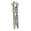
- Assembly
| Deposited unit | 
| ||||||||
|---|---|---|---|---|---|---|---|---|---|
| 1 | 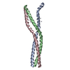
| ||||||||
| 2 | 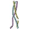
| ||||||||
| 3 | 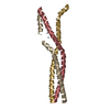
| ||||||||
| 4 | 
| ||||||||
| Unit cell |
|
-Components
| #1: Protein | Mass: 8269.400 Da / Num. of mol.: 12 / Fragment: oligomerization domain Source method: isolated from a genetically manipulated source Source: (gene. exp.) Zaire ebolavirus / Gene: VP35 / Plasmid: pET28 / Production host:  #2: Chemical | ChemComp-HG /   Mass: 200.590 Da / Num. of mol.: 11 / Source method: obtained synthetically / Formula: Hg Mass: 200.590 Da / Num. of mol.: 11 / Source method: obtained synthetically / Formula: Hg |
|---|
-Experimental details
-Experiment
| Experiment | Method: X-RAY DIFFRACTION / Number of used crystals: 1 |
|---|
- Sample preparation
| Crystal | Density Matthews: 3 Å3/Da / Density % sol: 58.94 % / Mosaicity: 0.25 ° |
|---|---|
| Crystal grow | Temperature: 296 K / Method: vapor diffusion / pH: 7.5 / Details: 50 mM HEPES-NaOH pH 7.5, 2.6 M Na-acetate |
-Data collection
| Diffraction | Mean temperature: 100 K | ||||||||||||||||||||||||||||||
|---|---|---|---|---|---|---|---|---|---|---|---|---|---|---|---|---|---|---|---|---|---|---|---|---|---|---|---|---|---|---|---|
| Diffraction source | Source: SYNCHROTRON / Site: ESRF  / Beamline: ID29 / Wavelength: 1.008 Å / Beamline: ID29 / Wavelength: 1.008 Å | ||||||||||||||||||||||||||||||
| Detector | Type: DECTRIS PILATUS 6M / Detector: PIXEL / Date: Jun 25, 2016 | ||||||||||||||||||||||||||||||
| Radiation | Protocol: SINGLE WAVELENGTH / Monochromatic (M) / Laue (L): M / Scattering type: x-ray | ||||||||||||||||||||||||||||||
| Radiation wavelength | Wavelength: 1.008 Å / Relative weight: 1 | ||||||||||||||||||||||||||||||
| Reflection | Resolution: 3.49→46.57 Å / Num. obs: 15726 / % possible obs: 99.2 % / Redundancy: 16.7 % / CC1/2: 0.998 / Rmerge(I) obs: 0.242 / Rpim(I) all: 0.06 / Rrim(I) all: 0.249 / Net I/σ(I): 10 / Num. measured all: 262316 / Scaling rejects: 1 | ||||||||||||||||||||||||||||||
| Reflection shell | Diffraction-ID: 1
|
-Phasing
| Phasing | Method: SAD |
|---|
- Processing
| Software |
| ||||||||||||||||||||||||||||||||||||||||||||||||||||||||||||
|---|---|---|---|---|---|---|---|---|---|---|---|---|---|---|---|---|---|---|---|---|---|---|---|---|---|---|---|---|---|---|---|---|---|---|---|---|---|---|---|---|---|---|---|---|---|---|---|---|---|---|---|---|---|---|---|---|---|---|---|---|---|
| Refinement | Method to determine structure: SAD / Resolution: 3.49→30 Å / Cor.coef. Fo:Fc: 0.935 / Cor.coef. Fo:Fc free: 0.888 / WRfactor Rfree: 0.2611 / WRfactor Rwork: 0.1987 / FOM work R set: 0.7975 / SU B: 34.358 / SU ML: 0.511 / SU Rfree: 0.6856 / Cross valid method: THROUGHOUT / σ(F): 0 / ESU R Free: 0.686 / Stereochemistry target values: MAXIMUM LIKELIHOOD Details: HYDROGENS HAVE BEEN ADDED IN THE RIDING POSITIONS U VALUES : REFINED INDIVIDUALLY
| ||||||||||||||||||||||||||||||||||||||||||||||||||||||||||||
| Solvent computation | Ion probe radii: 0.8 Å / Shrinkage radii: 0.8 Å / VDW probe radii: 1.2 Å / Solvent model: MASK | ||||||||||||||||||||||||||||||||||||||||||||||||||||||||||||
| Displacement parameters | Biso max: 238.74 Å2 / Biso mean: 99.538 Å2 / Biso min: 57.26 Å2
| ||||||||||||||||||||||||||||||||||||||||||||||||||||||||||||
| Refinement step | Cycle: final / Resolution: 3.49→30 Å
| ||||||||||||||||||||||||||||||||||||||||||||||||||||||||||||
| Refine LS restraints |
| ||||||||||||||||||||||||||||||||||||||||||||||||||||||||||||
| LS refinement shell | Resolution: 3.491→3.581 Å / Rfactor Rfree error: 0 / Total num. of bins used: 20
|
