 Movie
Movie Controller

[English] 日本語
 Yorodumi
- PDB-4njo: crystal structure of cofactor(NAD+) bound 3-phosphoglycerate dehy... -
+ Open data

- Basic information
| Entry | Database: PDB / ID: 4njo | ||||||
|---|---|---|---|---|---|---|---|
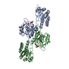
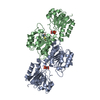
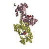
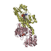
| Title | crystal structure of cofactor(NAD+) bound 3-phosphoglycerate dehydrogenase in Entamoeba histolytica | ||||||
 Components Components | D-3-phosphoglycerate dehydrogenase, putative | ||||||
 Keywords Keywords | OXIDOREDUCTASE / Rossmann Fold / Dehydrogenase | ||||||
| Function / homology |  Function and homology information Function and homology informationphosphoglycerate dehydrogenase / phosphoglycerate dehydrogenase activity / NAD binding / identical protein binding Similarity search - Function | ||||||
| Biological species |   Entamoeba histolytica (eukaryote) Entamoeba histolytica (eukaryote) | ||||||
| Method |  X-RAY DIFFRACTION / MOLECULAR REPLACEMENT / Resolution: 2.22 Å X-RAY DIFFRACTION / MOLECULAR REPLACEMENT / Resolution: 2.22 Å | ||||||
 Authors Authors | Singh, R.K. / Gourinath, S. | ||||||
 Citation Citation | Journal: Febs J. / Year: 2014 Title: Crystal structures and kinetics of Type III 3-phosphoglycerate dehydrogenase reveal catalysis by lysine. Authors: Singh, R.K. / Raj, I. / Pujari, R. / Gourinath, S. | ||||||
| History |
|
- Structure visualization
| Structure viewer | Molecule: MolmilJmol/JSmol |
|---|
- Downloads & links
-Download
| PDBx/mmCIF format | 4njo.cif.gz | 131.1 KB | Display | PDBx/mmCIF format |
|---|---|---|---|---|
| PDB format | pdb4njo.ent.gz | 102.2 KB | Display | PDB format |
| PDBx/mmJSON format | 4njo.json.gz | Tree view | PDBx/mmJSON format | |
| Others |  Other downloads Other downloads |
-Validation report
| Summary document | 4njo_validation.pdf.gz | 761.5 KB | Display | wwPDB validaton report |
|---|---|---|---|---|
| Full document | 4njo_full_validation.pdf.gz | 765.5 KB | Display | |
| Data in XML | 4njo_validation.xml.gz | 28 KB | Display | |
| Data in CIF | 4njo_validation.cif.gz | 36.7 KB | Display | |
| Arichive directory | https://data.pdbj.org/pub/pdb/validation_reports/nj/4njoftp://data.pdbj.org/pub/pdb/validation_reports/nj/4njo | HTTPS FTP |
-Related structure data

-Links
 PDBj
PDBj- Assembly
| Deposited unit | 
| ||||||||
|---|---|---|---|---|---|---|---|---|---|
| 1 |
| ||||||||
| Unit cell |
|
-Components
-Protein , 1 types, 2 molecules AB
| #1: Protein | Mass: 34749.449 Da / Num. of mol.: 2 Source method: isolated from a genetically manipulated source Source: (gene. exp.) Entamoeba histolytica (eukaryote) / Strain: HM1:IMSS / Gene: EhPGDH, EHI_060860 / Plasmid: pET21 / Production host:  |
|---|
-Non-polymers , 5 types, 153 molecules 


| #2: Chemical | ChemComp-NAD /  Mass: 663.425 Da / Num. of mol.: 1 / Source method: obtained synthetically / Formula: C21H27N7O14P2 / Comment: NAD*YM Mass: 663.425 Da / Num. of mol.: 1 / Source method: obtained synthetically / Formula: C21H27N7O14P2 / Comment: NAD*YM | ||||||
|---|---|---|---|---|---|---|---|
| #3: Chemical |  Mass: 46.025 Da / Num. of mol.: 2 / Source method: obtained synthetically / Formula: CH2O2 Mass: 46.025 Da / Num. of mol.: 2 / Source method: obtained synthetically / Formula: CH2O2#4: Chemical | ChemComp-TRS / |  Mass: 122.143 Da / Num. of mol.: 1 / Source method: obtained synthetically / Formula: C4H12NO3 / Comment: pH buffer*YM Mass: 122.143 Da / Num. of mol.: 1 / Source method: obtained synthetically / Formula: C4H12NO3 / Comment: pH buffer*YM#5: Chemical | ChemComp-GOL / |  Mass: 92.094 Da / Num. of mol.: 1 / Source method: obtained synthetically / Formula: C3H8O3 Mass: 92.094 Da / Num. of mol.: 1 / Source method: obtained synthetically / Formula: C3H8O3#6: Water | ChemComp-HOH / | Mass: 18.015 Da / Num. of mol.: 148 / Source method: isolated from a natural source / Formula: H2O |
-Experimental details
-Experiment
| Experiment | Method: X-RAY DIFFRACTION / Number of used crystals: 1 |
|---|
- Sample preparation
| Crystal | Density Matthews: 1.94 Å3/Da / Density % sol: 36.47 % |
|---|---|
| Crystal grow | Temperature: 289 K / Method: vapor diffusion, hanging drop / pH: 7.5 Details: 25% PEG 3350, 100mM Tris pH 7.0-8.0 300mM sodium formate, 5% glycerol , VAPOR DIFFUSION, HANGING DROP, temperature 289K |
-Data collection
| Diffraction | Mean temperature: 77 K |
|---|---|
| Diffraction source | Source: ROTATING ANODE / Type: RIGAKU MICROMAX-007 / Wavelength: 1.54 Å |
| Detector | Type: RIGAKU RAXIS IV++ / Detector: IMAGE PLATE / Date: Feb 15, 2012 |
| Radiation | Monochromator: Cu-Ni / Protocol: SINGLE WAVELENGTH / Monochromatic (M) / Laue (L): M / Scattering type: x-ray |
| Radiation wavelength | Wavelength: 1.54 Å / Relative weight: 1 |
| Reflection | Resolution: 2.22→52.09 Å / Num. obs: 24702 / % possible obs: 95.6 % / Observed criterion σ(F): 0 / Observed criterion σ(I): -3 |
| Reflection shell | Resolution: 2.22→2.3 Å / % possible all: 90.7 |
- Processing
| Software |
| ||||||||||||||||||||||||||||||||||||||||||||||||||||||||||||||||||||||||||||||||||||||||||||||||||||||||||||||||||||||||||||||||||||||||||||||||||||||||||||||||||||||||||||||||||||||
|---|---|---|---|---|---|---|---|---|---|---|---|---|---|---|---|---|---|---|---|---|---|---|---|---|---|---|---|---|---|---|---|---|---|---|---|---|---|---|---|---|---|---|---|---|---|---|---|---|---|---|---|---|---|---|---|---|---|---|---|---|---|---|---|---|---|---|---|---|---|---|---|---|---|---|---|---|---|---|---|---|---|---|---|---|---|---|---|---|---|---|---|---|---|---|---|---|---|---|---|---|---|---|---|---|---|---|---|---|---|---|---|---|---|---|---|---|---|---|---|---|---|---|---|---|---|---|---|---|---|---|---|---|---|---|---|---|---|---|---|---|---|---|---|---|---|---|---|---|---|---|---|---|---|---|---|---|---|---|---|---|---|---|---|---|---|---|---|---|---|---|---|---|---|---|---|---|---|---|---|---|---|---|---|
| Refinement | Method to determine structure: MOLECULAR REPLACEMENT / Resolution: 2.22→52.09 Å / Cor.coef. Fo:Fc: 0.953 / Cor.coef. Fo:Fc free: 0.929 / Occupancy max: 1 / Occupancy min: 0.4 / SU B: 7.397 / SU ML: 0.187 / SU R Cruickshank DPI: 0.5288 / Cross valid method: THROUGHOUT / ESU R: 0.532 / ESU R Free: 0.259 / Stereochemistry target values: MAXIMUM LIKELIHOOD / Details: HYDROGENS HAVE BEEN USED IF PRESENT IN THE INPUT
| ||||||||||||||||||||||||||||||||||||||||||||||||||||||||||||||||||||||||||||||||||||||||||||||||||||||||||||||||||||||||||||||||||||||||||||||||||||||||||||||||||||||||||||||||||||||
| Solvent computation | Ion probe radii: 0.8 Å / Shrinkage radii: 0.8 Å / VDW probe radii: 1.2 Å / Solvent model: MASK | ||||||||||||||||||||||||||||||||||||||||||||||||||||||||||||||||||||||||||||||||||||||||||||||||||||||||||||||||||||||||||||||||||||||||||||||||||||||||||||||||||||||||||||||||||||||
| Displacement parameters | Biso mean: 45.223 Å2
| ||||||||||||||||||||||||||||||||||||||||||||||||||||||||||||||||||||||||||||||||||||||||||||||||||||||||||||||||||||||||||||||||||||||||||||||||||||||||||||||||||||||||||||||||||||||
| Refinement step | Cycle: LAST / Resolution: 2.22→52.09 Å
| ||||||||||||||||||||||||||||||||||||||||||||||||||||||||||||||||||||||||||||||||||||||||||||||||||||||||||||||||||||||||||||||||||||||||||||||||||||||||||||||||||||||||||||||||||||||
| Refine LS restraints |
|