[English] 日本語
Yorodumi
- PDB-4nho: Structure of the spliceosomal DEAD-box protein Prp28 -

+
Open data


ID or keywords:

Loading...

-
Basic information

Entry
Database: PDB / ID: 4nho
TitleStructure of the spliceosomal DEAD-box protein Prp28
ComponentsProbable ATP-dependent RNA helicase DDX23
KeywordsHYDROLASE / DEAD-BOX / HELICASE
Function / homology
Function and homology information


R-loop processing / cis assembly of pre-catalytic spliceosome / RNA splicing, via transesterification reactions / mRNA Splicing - Minor Pathway / U5 snRNP / U4/U6 x U5 tri-snRNP complex / catalytic step 2 spliceosome / mRNA Splicing - Major Pathway / RNA splicing / mRNA splicing, via spliceosome ...R-loop processing / cis assembly of pre-catalytic spliceosome / RNA splicing, via transesterification reactions / mRNA Splicing - Minor Pathway / U5 snRNP / U4/U6 x U5 tri-snRNP complex / catalytic step 2 spliceosome / mRNA Splicing - Major Pathway / RNA splicing / mRNA splicing, via spliceosome / RNA helicase activity / RNA helicase / mRNA binding / chromatin / nucleolus / ATP hydrolysis activity / RNA binding / extracellular exosome / nucleoplasm / ATP binding / nucleus
Similarity search - Function
DEAD-box subfamily ATP-dependent helicases signature. / ATP-dependent RNA helicase DEAD-box, conserved site / RNA helicase, DEAD-box type, Q motif / DEAD-box RNA helicase Q motif profile. / DEAD/DEAH box helicase / DEAD/DEAH box helicase domain / Helicase conserved C-terminal domain / helicase superfamily c-terminal domain / Superfamilies 1 and 2 helicase C-terminal domain profile. / Superfamilies 1 and 2 helicase ATP-binding type-1 domain profile. ...DEAD-box subfamily ATP-dependent helicases signature. / ATP-dependent RNA helicase DEAD-box, conserved site / RNA helicase, DEAD-box type, Q motif / DEAD-box RNA helicase Q motif profile. / DEAD/DEAH box helicase / DEAD/DEAH box helicase domain / Helicase conserved C-terminal domain / helicase superfamily c-terminal domain / Superfamilies 1 and 2 helicase C-terminal domain profile. / Superfamilies 1 and 2 helicase ATP-binding type-1 domain profile. / DEAD-like helicases superfamily / Helicase, C-terminal / Helicase superfamily 1/2, ATP-binding domain / P-loop containing nucleotide triphosphate hydrolases / Rossmann fold / P-loop containing nucleoside triphosphate hydrolase / 3-Layer(aba) Sandwich / Alpha Beta
Similarity search - Domain/homology
: / Probable ATP-dependent RNA helicase DDX23
Similarity search - Component
Biological speciesHomo sapiens (human)
MethodX-RAY DIFFRACTION / SYNCHROTRON / SAD / Resolution: 2 Å
AuthorsMoehlmann, S. / Neumann, P. / Ficner, R.
CitationJournal: Acta Crystallogr.,Sect.D / Year: 2014
Title: Structural and functional analysis of the human spliceosomal DEAD-box helicase Prp28.
Authors: Mohlmann, S. / Mathew, R. / Neumann, P. / Schmitt, A. / Luhrmann, R. / Ficner, R.
History
DepositionNov 5, 2013Deposition site: RCSB / Processing site: RCSB
Revision 1.0Jun 18, 2014Provider: repository / Type: Initial release
Revision 1.1Sep 24, 2014Group: Database references
Revision 1.2Sep 4, 2019Group: Data collection / Derived calculations / Category: reflns / struct_conn
Item: _reflns.pdbx_Rsym_value / _struct_conn.pdbx_leaving_atom_flag
Revision 1.3Sep 20, 2023Group: Data collection / Database references ...Data collection / Database references / Derived calculations / Refinement description
Category: chem_comp_atom / chem_comp_bond ...chem_comp_atom / chem_comp_bond / database_2 / pdbx_initial_refinement_model / struct_ref_seq_dif / struct_site
Item: _database_2.pdbx_DOI / _database_2.pdbx_database_accession ..._database_2.pdbx_DOI / _database_2.pdbx_database_accession / _struct_ref_seq_dif.details / _struct_site.pdbx_auth_asym_id / _struct_site.pdbx_auth_comp_id / _struct_site.pdbx_auth_seq_id
Revision 1.4Dec 6, 2023Group: Data collection / Category: chem_comp_atom / chem_comp_bond / Item: _chem_comp_atom.atom_id / _chem_comp_bond.atom_id_2

-
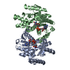
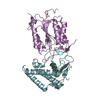
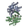
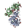
Structure visualization

Structure viewerMolecule:
MolmilJmol/JSmol

Downloads & links

-
Assembly

Deposited unit
A: Probable ATP-dependent RNA helicase DDX23
hetero molecules


Theoretical massNumber of molelcules
Total (without water)56,09910
Polymers55,0121
Non-polymers1,0869
Water3,963220
1


  • Idetical with deposited unit
  • defined by author
TypeNameSymmetry operationNumber
identity operation1_555x,y,z1
Unit cell
Length a, b, c (Å)125.430, 136.770, 73.220
Angle α, β, γ (deg.)90.000, 90.000, 90.000
Int Tables number20
Space group name H-MC2221
Components on special symmetry positions
IDModelComponents
11A-1199-

HOH

-
Components

-
Protein , 1 types, 1 molecules A

#1: Protein Probable ATP-dependent RNA helicase DDX23 / 100 kDa U5 snRNP-specific protein / DEAD box protein 23 / PRP28 homolog / U5-100kD


Mass: 55012.285 Da / Num. of mol.: 1 / Fragment: RecA domains (UNP residues 338-820)
Source method: isolated from a genetically manipulated source
Source: (gene. exp.) Homo sapiens (human) / Gene: DDX23 / Production host: Escherichia coli (E. coli) / Strain (production host): BL21 STAR (DE3) / References: UniProt: Q9BUQ8, RNA helicase

-
Non-polymers , 5 types, 229 molecules

#2: Chemical
ChemComp-SO4 / SULFATE ION


Mass: 96.063 Da / Num. of mol.: 5 / Source method: obtained synthetically / Formula: SO4
#3: Chemical ChemComp-GOL / GLYCEROL / GLYCERIN / PROPANE-1,2,3-TRIOL


Mass: 92.094 Da / Num. of mol.: 2 / Source method: obtained synthetically / Formula: C3H8O3
#4: Chemical ChemComp-CXS / 3-CYCLOHEXYL-1-PROPYLSULFONIC ACID


Mass: 221.317 Da / Num. of mol.: 1 / Source method: obtained synthetically / Formula: C9H19NO3S / Comment: pH buffer*YM
#5: Chemical ChemComp-HG / MERCURY (II) ION


Mass: 200.590 Da / Num. of mol.: 1 / Source method: obtained synthetically / Formula: Hg
#6: Water ChemComp-HOH / water


Mass: 18.015 Da / Num. of mol.: 220 / Source method: isolated from a natural source / Formula: H2O

-
Experimental details

-
Experiment

ExperimentMethod: X-RAY DIFFRACTION / Number of used crystals: 1

-
Sample preparation

CrystalDensity Matthews: 2.85 Å3/Da / Density % sol: 56.9 %
Crystal growTemperature: 298 K / Method: vapor diffusion, sitting drop / pH: 9
Details: 2.2 M ammonium sulfate, 20 % (v/v) glycerol, 0.1 M CAPS, mixing 1 ul of protein with 1 ul of reservoir solution, pH 9.0, VAPOR DIFFUSION, SITTING DROP, temperature 298K

-
Data collection

Diffraction
IDMean temperature (K)Crystal-ID
11001
21
Diffraction source
SourceSiteBeamlineIDWavelength (Å)
SYNCHROTRONBESSY 14.111.001212
SYNCHROTRONBESSY 14.121.00858
DetectorType: MARMOSAIC 225 mm CCD / Detector: CCD / Date: Jun 6, 2005 / Details: mirrors
RadiationMonochromator: GRAPHITE / Protocol: SINGLE WAVELENGTH / Monochromatic (M) / Laue (L): M / Scattering type: x-ray
Radiation wavelength
IDWavelength (Å)Relative weight
11.0012121
21.008581
ReflectionRedundancy: 4.9 % / Av σ(I) over netI: 6.7 / Number: 209358 / Rmerge(I) obs: 0.038 / Rsym value: 0.064 / D res high: 2 Å / Num. obs: 81386 / % possible obs: 99.2
Diffraction reflection shell

ID: 1

Highest resolution (Å)Lowest resolution (Å)Num. obs% possible obs (%)Rmerge(I) obsRsym valueRedundancy
5039.080.0290.0264.6
175011686.60.0130.054.9
141710096.20.010.0534.9
4.5514662398.70.0150.0425
4.124.55239197.90.0190.0665
3.694.12359298.50.0190.1065
3.263.695861990.0240.1525
2.43.262837599.50.0620.245
2.22.41407299.50.2050.3685
2.12.2915299.30.3310.5844.9
22.11110499.20.498
ReflectionResolution: 2→39.97 Å / Num. all: 42906 / Num. obs: 42906 / % possible obs: 100 % / Observed criterion σ(F): 0 / Observed criterion σ(I): 0 / Redundancy: 4.9 % / Biso Wilson estimate: 32.7 Å2 / Rmerge(I) obs: 0.064 / Net I/σ(I): 12.1
Reflection shell

Diffraction-ID: 1

Resolution (Å)Redundancy (%)Rmerge(I) obsMean I/σ(I) obsNum. unique allRsym value% possible all
2-2.114.90.5842.362190.652100
6.32-39.974.60.02630.214590.02999.1

-
Processing

Software
NameVersionClassificationNB
SCALA3.3.12data scaling
SOLOMONphasing
PHENIX1.5_2refinement
PDB_EXTRACT3.11data extraction
MAR345dtbdata collection
MOSFLMdata reduction
SHARPphasing
RefinementMethod to determine structure: SAD
Starting model: PDB entry 2DB3
Resolution: 2→28.825 Å / Occupancy max: 1 / Occupancy min: 0.23 / SU ML: 0.25 / Cross valid method: THROUGHOUT / σ(F): 1.34 / σ(I): 0 / Phase error: 26.89 / Stereochemistry target values: ML
RfactorNum. reflection% reflectionSelection details
Rfree0.2176 2159 5.04 %RANDOM
Rwork0.1931 ---
obs0.1943 42839 99.87 %-
all-42906 --
Solvent computationShrinkage radii: 0.8 Å / VDW probe radii: 1 Å / Solvent model: FLAT BULK SOLVENT MODEL / Bsol: 74.104 Å2 / ksol: 0.361 e/Å3
Displacement parametersBiso max: 187.48 Å2 / Biso mean: 59.0384 Å2 / Biso min: 26.43 Å2
Baniso -1Baniso -2Baniso -3
1-24.5474 Å2-0 Å2-0 Å2
2---11.2129 Å2-0 Å2
3----13.3345 Å2
Refinement stepCycle: LAST / Resolution: 2→28.825 Å
ProteinNucleic acidLigandSolventTotal
Num. atoms3505 0 52 220 3777
Refine LS restraints
Refine-IDTypeDev idealNumber
X-RAY DIFFRACTIONf_bond_d0.0123679
X-RAY DIFFRACTIONf_angle_d1.2764983
X-RAY DIFFRACTIONf_chiral_restr0.086551
X-RAY DIFFRACTIONf_plane_restr0.006643
X-RAY DIFFRACTIONf_dihedral_angle_d18.9181418
LS refinement shell

Refine-ID: X-RAY DIFFRACTION / Total num. of bins used: 15 / % reflection obs: 100 %

Resolution (Å)Rfactor RfreeNum. reflection RfreeRfactor RworkNum. reflection RworkNum. reflection all
2-2.04650.3971230.33826992822
2.0465-2.09770.3151520.296626902842
2.0977-2.15440.27581430.25726892832
2.1544-2.21780.30251590.229426592818
2.2178-2.28930.21711440.208426452789
2.2893-2.37110.24871400.185527042844
2.3711-2.4660.21151440.179327032847
2.466-2.57820.23071460.179126722818
2.5782-2.7140.23881310.191927152846
2.714-2.88390.22821490.194526992848
2.8839-3.10630.25831480.20227172865
3.1063-3.41850.21651250.184127262851
3.4185-3.91210.171650.167827262891
3.9121-4.92480.17491570.155427582915
4.9248-28.82830.19831330.194128783011
Refinement TLS params.

Method: refined / Refine-ID: X-RAY DIFFRACTION

IDL112)L122)L132)L222)L232)L332)S11 (Å °)S12 (Å °)S13 (Å °)S21 (Å °)S22 (Å °)S23 (Å °)S31 (Å °)S32 (Å °)S33 (Å °)T112)T122)T132)T222)T232)T332)Origin x (Å)Origin y (Å)Origin z (Å)
10.2456-0.1138-0.17240.782-0.20960.2354-0.22150.3160.1438-0.10980.06070.59020.3638-0.1980.19380.618-0.0724-0.08850.4173-0.13130.568716.174439.528919.0778
21.43750.32880.44461.56590.55681.1309-0.02480.16260.0327-0.6904-0.0928-0.0914-0.0858-0.04210.08670.53440.08420.08280.2530.01670.249440.191261.42418.6217
31.538-0.29460.0043.04010.40221.1499-0.0203-0.12050.01430.4607-0.2502-0.14620.157-0.13570.19460.508-0.08240.02560.35050.00430.337235.981646.33748.5969
40.20990.0008-0.14531.61671.52081.7613-0.1372-0.03230.09860.3536-0.74820.66690.851-1.03220.72170.8378-0.290.35930.8489-0.33190.778318.251540.84251.1186
Refinement TLS group
IDRefine-IDRefine TLS-IDSelection detailsAuth asym-IDAuth seq-ID
1X-RAY DIFFRACTION1CHAIN A AND (RESID 352:366)A345 - 366
2X-RAY DIFFRACTION2CHAIN A AND (RESID 367:632)A367 - 632
3X-RAY DIFFRACTION3CHAIN A AND (RESID 633:672 OR RESID 733:806)A633 - 672
4X-RAY DIFFRACTION4CHAIN A AND (RESID 673:732)A673 - 732

+
About Yorodumi

-
News

-
Feb 9, 2022. New format data for meta-information of EMDB entries

New format data for meta-information of EMDB entries

  • Version 3 of the EMDB header file is now the official format.
  • The previous official version 1.9 will be removed from the archive.

Related info.:EMDB header

External links:wwPDB to switch to version 3 of the EMDB data model

-
Aug 12, 2020. Covid-19 info

Covid-19 info

URL: https://pdbjlc1.pdbj.org/emnavi/covid19.php

New page: Covid-19 featured information page in EM Navigator.

Related info.:Covid-19 info / Mar 5, 2020. Novel coronavirus structure data

+
Mar 5, 2020. Novel coronavirus structure data

Novel coronavirus structure data

Related info.:Yorodumi Speices / Aug 12, 2020. Covid-19 info

External links:COVID-19 featured content - PDBj / Molecule of the Month (242):Coronavirus Proteases

+
Jan 31, 2019. EMDB accession codes are about to change! (news from PDBe EMDB page)

EMDB accession codes are about to change! (news from PDBe EMDB page)

  • The allocation of 4 digits for EMDB accession codes will soon come to an end. Whilst these codes will remain in use, new EMDB accession codes will include an additional digit and will expand incrementally as the available range of codes is exhausted. The current 4-digit format prefixed with “EMD-” (i.e. EMD-XXXX) will advance to a 5-digit format (i.e. EMD-XXXXX), and so on. It is currently estimated that the 4-digit codes will be depleted around Spring 2019, at which point the 5-digit format will come into force.
  • The EM Navigator/Yorodumi systems omit the EMD- prefix.

Related info.:Q: What is EMD? / ID/Accession-code notation in Yorodumi/EM Navigator

External links:EMDB Accession Codes are Changing Soon! / Contact to PDBj

+
Jul 12, 2017. Major update of PDB

Major update of PDB

  • wwPDB released updated PDB data conforming to the new PDBx/mmCIF dictionary.
  • This is a major update changing the version number from 4 to 5, and with Remediation, in which all the entries are updated.
  • In this update, many items about electron microscopy experimental information are reorganized (e.g. em_software).
  • Now, EM Navigator and Yorodumi are based on the updated data.

External links:wwPDB Remediation / Enriched Model Files Conforming to OneDep Data Standards Now Available in the PDB FTP Archive

-
Yorodumi

Thousand views of thousand structures

  • Yorodumi is a browser for structure data from EMDB, PDB, SASBDB, etc.
  • This page is also the successor to EM Navigator detail page, and also detail information page/front-end page for Omokage search.
  • The word "yorodu" (or yorozu) is an old Japanese word meaning "ten thousand". "mi" (miru) is to see.

Related info.:EMDB / PDB / SASBDB / Comparison of 3 databanks / Yorodumi Search / Aug 31, 2016. New EM Navigator & Yorodumi / Yorodumi Papers / Jmol/JSmol / Function and homology information / Changes in new EM Navigator and Yorodumi

Read more