 Movie
Movie Controller

[English] 日本語
 Yorodumi
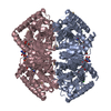
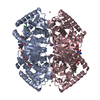
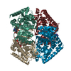
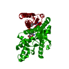
- PDB-1uxi: Large improvement in the thermal stability of a tetrameric malate... -
+ Open data

- Basic information
| Entry | Database: PDB / ID: 1uxi | |||||||||
|---|---|---|---|---|---|---|---|---|---|---|
| Title | Large improvement in the thermal stability of a tetrameric malate dehydrogenase by single point mutations at the dimer-dimer interface | |||||||||
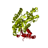
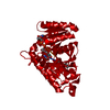
 Components Components | MALATE DEHYDROGENASE | |||||||||
 Keywords Keywords | OXIDOREDUCTASE / TRICARBOXYLIC ACID CYCLE / MALATE DEHYDROGENASE | |||||||||
| Function / homology |  Function and homology information Function and homology information(S)-malate dehydrogenase (NAD+, oxaloacetate-forming) / L-malate dehydrogenase (NAD+) activity / carboxylic acid metabolic process / tricarboxylic acid cycle / cytoplasm Similarity search - Function | |||||||||
| Biological species |   CHLOROFLEXUS AURANTIACUS (bacteria) CHLOROFLEXUS AURANTIACUS (bacteria) | |||||||||
| Method |  X-RAY DIFFRACTION / SYNCHROTRON / MOLECULAR REPLACEMENT / Resolution: 2.1 Å X-RAY DIFFRACTION / SYNCHROTRON / MOLECULAR REPLACEMENT / Resolution: 2.1 Å | |||||||||
 Authors Authors | Bjork, A. / Dalhus, B. / Mantzilas, D. / Eijsink, V.G.H. / Sirevag, R. | |||||||||
 Citation Citation | Journal: J.Mol.Biol. / Year: 2004 Title: Large Improvement in the Thermal Stability of a Tetrameric Malate Dehydrogenase by Single Point Mutations at the Dimer-Dimer Interface. Authors: Bjork, A. / Dalhus, B. / Mantzilas, D. / Sirevag, R. / Eijsink, V.G.H. | |||||||||
| History |
|
- Structure visualization
| Structure viewer | Molecule: MolmilJmol/JSmol |
|---|
- Downloads & links
-Download
| PDBx/mmCIF format | 1uxi.cif.gz | 138.8 KB | Display | PDBx/mmCIF format |
|---|---|---|---|---|
| PDB format | pdb1uxi.ent.gz | 107.9 KB | Display | PDB format |
| PDBx/mmJSON format | 1uxi.json.gz | Tree view | PDBx/mmJSON format | |
| Others |  Other downloads Other downloads |
-Validation report
| Arichive directory | https://data.pdbj.org/pub/pdb/validation_reports/ux/1uxiftp://data.pdbj.org/pub/pdb/validation_reports/ux/1uxi | HTTPS FTP |
|---|
-Related structure data
| Related structure data |  1uxgC  1uxhC  1uxjC  1uxkC 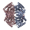 1guyS S: Starting model for refinement C: citing same article ( |
|---|---|
| Similar structure data |


-Links
 PDBj
PDBj


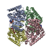
- Assembly
| Deposited unit | 
| |||||||||
|---|---|---|---|---|---|---|---|---|---|---|
| 1 | 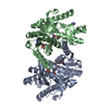
| |||||||||
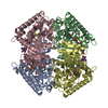
| Unit cell |
| |||||||||
| Components on special symmetry positions |
|
-Components
| #1: Protein | Mass: 32747.811 Da / Num. of mol.: 2 / Mutation: YES Source method: isolated from a genetically manipulated source Source: (gene. exp.) CHLOROFLEXUS AURANTIACUS (bacteria) / Production host: References: UniProt: P80040, (S)-malate dehydrogenase (NAD+, oxaloacetate-forming) #2: Chemical |   Mass: 663.425 Da / Num. of mol.: 2 / Source method: obtained synthetically / Formula: C21H27N7O14P2 / Comment: NAD*YM Mass: 663.425 Da / Num. of mol.: 2 / Source method: obtained synthetically / Formula: C21H27N7O14P2 / Comment: NAD*YM#3: Chemical | 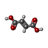  Mass: 116.072 Da / Num. of mol.: 2 / Source method: obtained synthetically / Formula: C4H4O4 Mass: 116.072 Da / Num. of mol.: 2 / Source method: obtained synthetically / Formula: C4H4O4#4: Chemical | ChemComp-NA / |   Mass: 22.990 Da / Num. of mol.: 1 / Source method: obtained synthetically / Formula: Na Mass: 22.990 Da / Num. of mol.: 1 / Source method: obtained synthetically / Formula: Na#5: Water | ChemComp-HOH / |  Mass: 18.015 Da / Num. of mol.: 450 / Source method: isolated from a natural source / Formula: H2O Mass: 18.015 Da / Num. of mol.: 450 / Source method: isolated from a natural source / Formula: H2OCompound details | ENGINEERED | |
|---|
-Experimental details
-Experiment
| Experiment | Method: X-RAY DIFFRACTION / Number of used crystals: 1 |
|---|
- Sample preparation
| Crystal | Density Matthews: 4.85 Å3/Da / Density % sol: 74 % |
|---|---|
| Crystal grow | pH: 6.5 / Details: 20-40% PEG400, 100 MM NASUCCINATE, PH 6.5 |
-Data collection
| Diffraction | Mean temperature: 100 K |
|---|---|
| Diffraction source | Source: SYNCHROTRON / Site: EMBL/DESY, HAMBURG  / Beamline: X31 / Wavelength: 1 / Beamline: X31 / Wavelength: 1 |
| Detector | Type: MARRESEARCH / Detector: IMAGE PLATE |
| Radiation | Protocol: SINGLE WAVELENGTH / Monochromatic (M) / Laue (L): M / Scattering type: x-ray |
| Radiation wavelength | Wavelength: 1 Å / Relative weight: 1 |
| Reflection | Resolution: 2.1→25 Å / Num. obs: 74855 / % possible obs: 91.4 % / Redundancy: 5.4 % / Rmerge(I) obs: 0.089 / Net I/σ(I): 20.5 |
| Reflection shell | Resolution: 2.1→2.18 Å / Rmerge(I) obs: 0.53 / Mean I/σ(I) obs: 3.7 / % possible all: 83.5 |
- Processing
| Software |
| ||||||||||||||||||||||||||||||||||||||||||||||||||||||||||||
|---|---|---|---|---|---|---|---|---|---|---|---|---|---|---|---|---|---|---|---|---|---|---|---|---|---|---|---|---|---|---|---|---|---|---|---|---|---|---|---|---|---|---|---|---|---|---|---|---|---|---|---|---|---|---|---|---|---|---|---|---|---|
| Refinement | Method to determine structure: MOLECULAR REPLACEMENT Starting model: PDB ENTRY 1GUY Resolution: 2.1→25 Å / Cross valid method: THROUGHOUT / σ(F): 2
| ||||||||||||||||||||||||||||||||||||||||||||||||||||||||||||
| Solvent computation | Solvent model: FLAT MODEL / Bsol: 66.7 Å2 | ||||||||||||||||||||||||||||||||||||||||||||||||||||||||||||
| Displacement parameters | Biso mean: 28.7 Å2 | ||||||||||||||||||||||||||||||||||||||||||||||||||||||||||||
| Refine analyze |
| ||||||||||||||||||||||||||||||||||||||||||||||||||||||||||||
| Refinement step | Cycle: LAST / Resolution: 2.1→25 Å
| ||||||||||||||||||||||||||||||||||||||||||||||||||||||||||||
| Refine LS restraints |
| ||||||||||||||||||||||||||||||||||||||||||||||||||||||||||||
| LS refinement shell | Resolution: 2.1→2.18 Å / Total num. of bins used: 10
|
