 Movie
Movie Controller

[English] 日本語
 Yorodumi
- PDB-4plz: Crystal structure of Plasmodium falciparum lactate dehydrogenase ... -
+ Open data

- Basic information
| Entry | Database: PDB / ID: 4plz | ||||||||||||
|---|---|---|---|---|---|---|---|---|---|---|---|---|---|
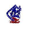
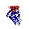
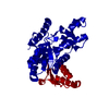
| Title | Crystal structure of Plasmodium falciparum lactate dehydrogenase mutant W107fA. | ||||||||||||
 Components Components | L-lactate dehydrogenase | ||||||||||||
 Keywords Keywords | OXIDOREDUCTASE / Ancestral Sequence Reconstruction / Dehydrogenase / Apicomplexa / Specificity | ||||||||||||
| Function / homology |  Function and homology information Function and homology informationL-lactate dehydrogenase / L-lactate dehydrogenase (NAD+) activity / lactate metabolic process / metal ion binding Similarity search - Function | ||||||||||||
| Biological species |  | ||||||||||||
| Method |  X-RAY DIFFRACTION / SYNCHROTRON / MOLECULAR REPLACEMENT / Resolution: 1.05 Å X-RAY DIFFRACTION / SYNCHROTRON / MOLECULAR REPLACEMENT / Resolution: 1.05 Å | ||||||||||||
 Authors Authors | Boucher, J.I. / Jacobowitz, J.R. / Beckett, B.C. / Classen, S. / Theobald, D.L. | ||||||||||||
| Funding support |  United States, 3items United States, 3items
| ||||||||||||
 Citation Citation | Journal: Elife / Year: 2014 Title: An atomic-resolution view of neofunctionalization in the evolution of apicomplexan lactate dehydrogenases. Authors: Boucher, J.I. / Jacobowitz, J.R. / Beckett, B.C. / Classen, S. / Theobald, D.L. | ||||||||||||
| History |
|
- Structure visualization
| Structure viewer | Molecule: MolmilJmol/JSmol |
|---|
- Downloads & links
-Download
| PDBx/mmCIF format | 4plz.cif.gz | 147 KB | Display | PDBx/mmCIF format |
|---|---|---|---|---|
| PDB format | pdb4plz.ent.gz | 112 KB | Display | PDB format |
| PDBx/mmJSON format | 4plz.json.gz | Tree view | PDBx/mmJSON format | |
| Others |  Other downloads Other downloads |
-Validation report
| Arichive directory | https://data.pdbj.org/pub/pdb/validation_reports/pl/4plzftp://data.pdbj.org/pub/pdb/validation_reports/pl/4plz | HTTPS FTP |
|---|
-Related structure data
| Related structure data |  4plcC  4plfC  4plgC  4plhC  4pltC  4plvC  4plwC  4plyC  1t2dS C: citing same article ( S: Starting model for refinement |
|---|---|
| Similar structure data |
-Links
 PDBj
PDBj


- Assembly
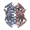
| Deposited unit | 
| ||||||||||||
|---|---|---|---|---|---|---|---|---|---|---|---|---|---|
| 1 | 
| ||||||||||||
| Unit cell |
| ||||||||||||
| Components on special symmetry positions |
|
-Components
| #1: Protein | Mass: 34862.453 Da / Num. of mol.: 1 Source method: isolated from a genetically manipulated source Source: (gene. exp.) Gene: LDH-P, LDH, ldh / Production host:  References: UniProt: Q71T02, UniProt: Q27743*PLUS, L-lactate dehydrogenase |
|---|---|
| #2: Chemical | ChemComp-OXM /   Mass: 89.050 Da / Num. of mol.: 1 / Source method: obtained synthetically / Formula: C2H3NO3 Mass: 89.050 Da / Num. of mol.: 1 / Source method: obtained synthetically / Formula: C2H3NO3 |
| #3: Chemical | ChemComp-NAI /   Mass: 665.441 Da / Num. of mol.: 1 / Source method: obtained synthetically / Formula: C21H29N7O14P2 Mass: 665.441 Da / Num. of mol.: 1 / Source method: obtained synthetically / Formula: C21H29N7O14P2 |
| #4: Water | ChemComp-HOH /  Mass: 18.015 Da / Num. of mol.: 547 / Source method: isolated from a natural source / Formula: H2O Mass: 18.015 Da / Num. of mol.: 547 / Source method: isolated from a natural source / Formula: H2O |
-Experimental details
-Experiment
| Experiment | Method: X-RAY DIFFRACTION / Number of used crystals: 1 |
|---|
- Sample preparation
| Crystal | Density Matthews: 2.27 Å3/Da / Density % sol: 45.84 % |
|---|---|
| Crystal grow | Temperature: 293 K / Method: vapor diffusion, hanging drop / pH: 7.5 Details: 20mg/mL protein with 1.2mM oxamate/2mM NADH; 22% PEG-1000 |
-Data collection
| Diffraction | Mean temperature: 90 K |
|---|---|
| Diffraction source | Source: SYNCHROTRON / Site: ALS / Beamline: 12.3.1 / Wavelength: 1.115866 Å |
| Detector | Type: ADSC QUANTUM 315r / Detector: CCD / Date: Dec 18, 2012 |
| Radiation | Monochromator: Si(111) / Protocol: SINGLE WAVELENGTH / Monochromatic (M) / Laue (L): M / Scattering type: x-ray |
| Radiation wavelength | Wavelength: 1.115866 Å / Relative weight: 1 |
| Reflection | Resolution: 1.05→50 Å / Num. obs: 284505 / % possible obs: 99.3 % / Redundancy: 2.9 % / Biso Wilson estimate: 9.15 Å2 / Rmerge(I) obs: 0.029 / Net I/σ(I): 18.9 |
| Reflection shell | Resolution: 1.05→1.08 Å / Redundancy: 1.5 % / Rmerge(I) obs: 0.42 / Mean I/σ(I) obs: 1.81 / % possible all: 97.1 |
- Processing
| Software |
| |||||||||||||||||||||||||||||||||||||||||||||||||||||||||||||||||||||||||||||||||||||||||||||||||||||||||||||||||||||||||||||||||||||||||||||||||||||||||||||||||||||||||||||||||||||||||||||||||||||||||||
|---|---|---|---|---|---|---|---|---|---|---|---|---|---|---|---|---|---|---|---|---|---|---|---|---|---|---|---|---|---|---|---|---|---|---|---|---|---|---|---|---|---|---|---|---|---|---|---|---|---|---|---|---|---|---|---|---|---|---|---|---|---|---|---|---|---|---|---|---|---|---|---|---|---|---|---|---|---|---|---|---|---|---|---|---|---|---|---|---|---|---|---|---|---|---|---|---|---|---|---|---|---|---|---|---|---|---|---|---|---|---|---|---|---|---|---|---|---|---|---|---|---|---|---|---|---|---|---|---|---|---|---|---|---|---|---|---|---|---|---|---|---|---|---|---|---|---|---|---|---|---|---|---|---|---|---|---|---|---|---|---|---|---|---|---|---|---|---|---|---|---|---|---|---|---|---|---|---|---|---|---|---|---|---|---|---|---|---|---|---|---|---|---|---|---|---|---|---|---|---|---|---|---|---|---|
| Refinement | Method to determine structure: MOLECULAR REPLACEMENT Starting model: 1T2D Resolution: 1.05→45.72 Å / SU ML: 0.06 / Cross valid method: FREE R-VALUE / σ(F): 1.35 / Phase error: 12.35 / Stereochemistry target values: ML
| |||||||||||||||||||||||||||||||||||||||||||||||||||||||||||||||||||||||||||||||||||||||||||||||||||||||||||||||||||||||||||||||||||||||||||||||||||||||||||||||||||||||||||||||||||||||||||||||||||||||||||
| Solvent computation | Shrinkage radii: 0.9 Å / VDW probe radii: 1.11 Å / Solvent model: FLAT BULK SOLVENT MODEL | |||||||||||||||||||||||||||||||||||||||||||||||||||||||||||||||||||||||||||||||||||||||||||||||||||||||||||||||||||||||||||||||||||||||||||||||||||||||||||||||||||||||||||||||||||||||||||||||||||||||||||
| Displacement parameters | Biso max: 57.92 Å2 / Biso mean: 14.663 Å2 / Biso min: 5.78 Å2 | |||||||||||||||||||||||||||||||||||||||||||||||||||||||||||||||||||||||||||||||||||||||||||||||||||||||||||||||||||||||||||||||||||||||||||||||||||||||||||||||||||||||||||||||||||||||||||||||||||||||||||
| Refinement step | Cycle: final / Resolution: 1.05→45.72 Å
| |||||||||||||||||||||||||||||||||||||||||||||||||||||||||||||||||||||||||||||||||||||||||||||||||||||||||||||||||||||||||||||||||||||||||||||||||||||||||||||||||||||||||||||||||||||||||||||||||||||||||||
| Refine LS restraints |
| |||||||||||||||||||||||||||||||||||||||||||||||||||||||||||||||||||||||||||||||||||||||||||||||||||||||||||||||||||||||||||||||||||||||||||||||||||||||||||||||||||||||||||||||||||||||||||||||||||||||||||
| LS refinement shell | Refine-ID: X-RAY DIFFRACTION / Total num. of bins used: 28
|
