 Movie
Movie Controller


+ Open data

- Basic information
| Entry | Database: PDB / ID: 4nfb | ||||||
|---|---|---|---|---|---|---|---|
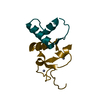
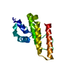
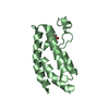
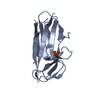
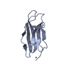
| Title | Structure of paired immunoglobulin-like type 2 receptor (PILR ) | ||||||
 Components Components | Paired immunoglobulin-like type 2 receptor alpha | ||||||
 Keywords Keywords | IMMUNE SYSTEM / IgV-like / immune-related inhibition receptor / HSV-1 gB / cell surface | ||||||
| Function / homology |  Function and homology information Function and homology informationMHC class I protein binding / Immunoregulatory interactions between a Lymphoid and a non-Lymphoid cell / signal transduction / extracellular exosome / plasma membrane Similarity search - Function | ||||||
| Biological species |  Homo sapiens (human) Homo sapiens (human) | ||||||
| Method |  X-RAY DIFFRACTION / SYNCHROTRON / MOLECULAR REPLACEMENT / Resolution: 1.6 Å X-RAY DIFFRACTION / SYNCHROTRON / MOLECULAR REPLACEMENT / Resolution: 1.6 Å | ||||||
 Authors Authors | Lu, Q. / Lu, G. / Qi, J. / Li, Y. / Zhang, Y. / Wang, H. / Fan, Z. / Yan, J. / Gao, G. | ||||||
 Citation Citation | Journal: Proc.Natl.Acad.Sci.USA / Year: 2014 Title: PILR alpha and PILR beta have a siglec fold and provide the basis of binding to sialic acid Authors: Lu, Q. / Lu, G. / Qi, J. / Wang, H. / Xuan, Y. / Wang, Q. / Li, Y. / Zhang, Y. / Zheng, C. / Fan, Z. / Yan, J. / Gao, G.F. | ||||||
| History |
|
- Structure visualization
| Structure viewer | Molecule: MolmilJmol/JSmol |
|---|
- Downloads & links
-Download
| PDBx/mmCIF format | 4nfb.cif.gz | 67 KB | Display | PDBx/mmCIF format |
|---|---|---|---|---|
| PDB format | pdb4nfb.ent.gz | 49.6 KB | Display | PDB format |
| PDBx/mmJSON format | 4nfb.json.gz | Tree view | PDBx/mmJSON format | |
| Others |  Other downloads Other downloads |
-Validation report
| Summary document | 4nfb_validation.pdf.gz | 422 KB | Display | wwPDB validaton report |
|---|---|---|---|---|
| Full document | 4nfb_full_validation.pdf.gz | 422.7 KB | Display | |
| Data in XML | 4nfb_validation.xml.gz | 8.4 KB | Display | |
| Data in CIF | 4nfb_validation.cif.gz | 11.3 KB | Display | |
| Arichive directory | https://data.pdbj.org/pub/pdb/validation_reports/nf/4nfbftp://data.pdbj.org/pub/pdb/validation_reports/nf/4nfb | HTTPS FTP |
-Related structure data


-Links
 PDBj
PDBj- Assembly
| Deposited unit | 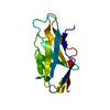
| ||||||||
|---|---|---|---|---|---|---|---|---|---|
| 1 |
| ||||||||
| Unit cell |
|
-Components
| #1: Protein | Mass: 14067.840 Da / Num. of mol.: 1 / Fragment: UNP residues 32-150 Source method: isolated from a genetically manipulated source Source: (gene. exp.) Homo sapiens (human) / Gene: PILR, PILRA / Plasmid: pET-21a / Production host:  |
|---|---|
| #2: Water | ChemComp-HOH /  Mass: 18.015 Da / Num. of mol.: 130 / Source method: isolated from a natural source / Formula: H2O Mass: 18.015 Da / Num. of mol.: 130 / Source method: isolated from a natural source / Formula: H2O |
-Experimental details
-Experiment
| Experiment | Method: X-RAY DIFFRACTION / Number of used crystals: 1 |
|---|
- Sample preparation
| Crystal | Density Matthews: 1.83 Å3/Da / Density % sol: 32.64 % |
|---|---|
| Crystal grow | Temperature: 277 K / Method: vapor diffusion, sitting drop / pH: 5.6 Details: 0.1M sodium citrate tribasic dehydrate pH 5.6, 20%(v/v) 2-propanol, 20%(w/v) polyethylene glycol 4,000, VAPOR DIFFUSION, SITTING DROP, temperature 277K |
-Data collection
| Diffraction | Mean temperature: 100 K |
|---|---|
| Diffraction source | Source: SYNCHROTRON / Site: SSRF  / Beamline: BL17U / Wavelength: 1 Å / Beamline: BL17U / Wavelength: 1 Å |
| Detector | Type: ADSC QUANTUM 315r / Detector: CCD / Date: Apr 10, 2011 |
| Radiation | Monochromator: GRAPHITE / Protocol: SINGLE WAVELENGTH / Monochromatic (M) / Laue (L): M / Scattering type: x-ray |
| Radiation wavelength | Wavelength: 1 Å / Relative weight: 1 |
| Reflection | Resolution: 1.6→50 Å / Num. all: 14159 / Num. obs: 13899 / % possible obs: 98.2 % / Observed criterion σ(F): 0 / Observed criterion σ(I): -3 / Redundancy: 7.7 % / Rmerge(I) obs: 0.06 / Rsym value: 0.06 / Net I/σ(I): 29.8 |
| Reflection shell | Resolution: 1.6→1.66 Å / Redundancy: 6.9 % / Rmerge(I) obs: 0.28 / Mean I/σ(I) obs: 5.92 / Num. unique all: 1289 / Rsym value: 0.28 / % possible all: 92.8 |
- Processing
| Software |
| ||||||||||||||||||||||||||||||||||||||||
|---|---|---|---|---|---|---|---|---|---|---|---|---|---|---|---|---|---|---|---|---|---|---|---|---|---|---|---|---|---|---|---|---|---|---|---|---|---|---|---|---|---|
| Refinement | Method to determine structure: MOLECULAR REPLACEMENT / Resolution: 1.6→33.07 Å / SU ML: 0.2 / σ(F): 0.13 / Phase error: 20.68 / Stereochemistry target values: ML Details: The starting model is obtained by single isomorphous replacement with a iodine derivative data.
| ||||||||||||||||||||||||||||||||||||||||
| Solvent computation | Shrinkage radii: 0.9 Å / VDW probe radii: 1.11 Å / Solvent model: FLAT BULK SOLVENT MODEL / Bsol: 59.577 Å2 / ksol: 0.416 e/Å3 | ||||||||||||||||||||||||||||||||||||||||
| Displacement parameters |
| ||||||||||||||||||||||||||||||||||||||||
| Refinement step | Cycle: LAST / Resolution: 1.6→33.07 Å
| ||||||||||||||||||||||||||||||||||||||||
| Refine LS restraints |
| ||||||||||||||||||||||||||||||||||||||||
| LS refinement shell | Refine-ID: X-RAY DIFFRACTION / Total num. of bins used: 5
| ||||||||||||||||||||||||||||||||||||||||
| Refinement TLS params. | Method: refined / Origin x: 10.198 Å / Origin y: -8.3398 Å / Origin z: -6.4539 Å
| ||||||||||||||||||||||||||||||||||||||||
| Refinement TLS group | Selection details: all |
