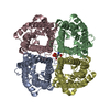
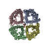
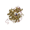
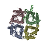
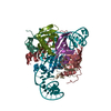
- PDB-4nef: X-ray structure of human Aquaporin 2 -
+
Open data
ID or keywords:
Loading...
-
Basic information
Entry
Database: PDB / ID: 4nef
Title
X-ray structure of human Aquaporin 2
Components
Aquaporin-2
Keywords
MEMBRANE PROTEIN / water channel / cadmium binding
Function / homology
Function and homology information
cellular response to water deprivation / renal water transport / glycerol transmembrane transporter activity / water transmembrane transporter activity / lumenal side of membrane / Passive transport by Aquaporins / glycerol transmembrane transport / cellular response to mercury ion / water channel activity / water transport ...cellular response to water deprivation / renal water transport / glycerol transmembrane transporter activity / water transmembrane transporter activity / lumenal side of membrane / Passive transport by Aquaporins / glycerol transmembrane transport / cellular response to mercury ion / water channel activity / water transport / metanephric collecting duct development / transport vesicle membrane / renal water homeostasis / cellular response to copper ion / actin filament organization / recycling endosome / Vasopressin regulates renal water homeostasis via Aquaporins / protein homotetramerization / basolateral plasma membrane / apical plasma membrane / perinuclear region of cytoplasm / Golgi apparatus / extracellular exosome / membrane / plasma membrane Similarity search - Function
Glycerol uptake facilitator protein / Glycerol uptake facilitator protein. / Aquaporin transporter / Major intrinsic protein, conserved site / MIP family signature. / Major intrinsic protein / Major intrinsic protein / Aquaporin-like / Up-down Bundle / Mainly Alpha Similarity search - Domain/homology
Aquaporin-2 / AQP-2 / ADH water channel / Aquaporin-CD / AQP-CD / Collecting duct water channel protein / WCH-CD ...AQP-2 / ADH water channel / Aquaporin-CD / AQP-CD / Collecting duct water channel protein / WCH-CD / Water channel protein for renal collecting duct
Mass: 25236.336 Da / Num. of mol.: 4 / Fragment: UNP residues 1-241 / Mutation: W2S Source method: isolated from a genetically manipulated source Source: (gene. exp.) Homo sapiens (human) / Gene: AQP2 / Production host: Pichia pastoris (fungus) / References: UniProt: P41181
In the structure databanks used in Yorodumi, some data are registered as the other names, "COVID-19 virus" and "2019-nCoV". Here are the details of the virus and the list of structure data.
Jan 31, 2019. EMDB accession codes are about to change! (news from PDBe EMDB page)
EMDB accession codes are about to change! (news from PDBe EMDB page)
The allocation of 4 digits for EMDB accession codes will soon come to an end. Whilst these codes will remain in use, new EMDB accession codes will include an additional digit and will expand incrementally as the available range of codes is exhausted. The current 4-digit format prefixed with “EMD-” (i.e. EMD-XXXX) will advance to a 5-digit format (i.e. EMD-XXXXX), and so on. It is currently estimated that the 4-digit codes will be depleted around Spring 2019, at which point the 5-digit format will come into force.
The EM Navigator/Yorodumi systems omit the EMD- prefix.
Related info.:Q: What is EMD? / ID/Accession-code notation in Yorodumi/EM Navigator
Yorodumi is a browser for structure data from EMDB, PDB, SASBDB, etc.
This page is also the successor to EM Navigator detail page, and also detail information page/front-end page for Omokage search.
The word "yorodu" (or yorozu) is an old Japanese word meaning "ten thousand". "mi" (miru) is to see.
Related info.:EMDB / PDB / SASBDB / Comparison of 3 databanks / Yorodumi Search / Aug 31, 2016. New EM Navigator & Yorodumi / Yorodumi Papers / Jmol/JSmol / Function and homology information / Changes in new EM Navigator and Yorodumi
 Movie
Movie Controller


 Open data
 Basic information
 Components
 Keywords
 Function and homology information
 Homo sapiens (human)
 X-RAY DIFFRACTION /
  Authors
 Citation
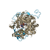
 Structure visualization
 Downloads & links
 Other downloads
 PDBj
PDBj

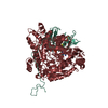
 Assembly
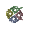
 Pichia pastoris (fungus) / References: UniProt: P41181
 Mass: 112.411 Da / Num. of mol.: 2 / Source method: obtained synthetically / Formula: Cd
 Mass: 65.409 Da / Num. of mol.: 1 / Source method: obtained synthetically / Formula: Zn
 Mass: 18.015 Da / Num. of mol.: 59 / Source method: isolated from a natural source / Formula: H2O
 Sample preparation
 / Beamline: ID14-4 / Wavelength: 0.9393
  Processing