 Movie
Movie Controller


+ Open data

- Basic information
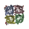
| Entry | Database: PDB / ID: 3d9s | ||||||
|---|---|---|---|---|---|---|---|
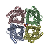
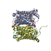
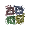
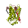
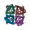
| Title | Human Aquaporin 5 (AQP5) - High Resolution X-ray Structure | ||||||
 Components Components | Aquaporin-5 | ||||||
 Keywords Keywords | MEMBRANE PROTEIN / Aquaporin / AQP / Aquaglyceroporin / water transport / lipid / Phosphatidylserine / PSF / NPA / ar/R / Water channel / Glycoprotein / Membrane / Transmembrane / Transport | ||||||
| Function / homology |  Function and homology information Function and homology informationPassive transport by Aquaporins / pancreatic juice secretion / saliva secretion / water channel activity / cellular hypotonic response / camera-type eye morphogenesis / water transport / odontogenesis / microvillus / basal plasma membrane ...Passive transport by Aquaporins / pancreatic juice secretion / saliva secretion / water channel activity / cellular hypotonic response / camera-type eye morphogenesis / water transport / odontogenesis / microvillus / basal plasma membrane / cytoplasmic vesicle membrane / carbon dioxide transport / protein homotetramerization / apical plasma membrane / endoplasmic reticulum / extracellular exosome / identical protein binding / plasma membrane Similarity search - Function | ||||||
| Biological species |  Homo sapiens (human) Homo sapiens (human) | ||||||
| Method |  X-RAY DIFFRACTION / SYNCHROTRON / AB INITIO / Resolution: 2 Å X-RAY DIFFRACTION / SYNCHROTRON / AB INITIO / Resolution: 2 Å | ||||||
 Authors Authors | Horsefield, R. / Norden, K. / Fellert, M. / Backmark, A. / Tornroth-Horsefield, S. / Terwisscha Van Scheltinga, A.C. / Kvassman, J. / Kjellbom, P. / Johanson, U. / Neutze, R. | ||||||
 Citation Citation | Journal: Proc.Natl.Acad.Sci.Usa / Year: 2008 Title: High-resolution x-ray structure of human aquaporin 5 Authors: Horsefield, R. / Norden, K. / Fellert, M. / Backmark, A. / Tornroth-Horsefield, S. / Terwisscha van Scheltinga, A.C. / Kvassman, J. / Kjellbom, P. / Johanson, U. / Neutze, R. | ||||||
| History |
|
- Structure visualization
| Structure viewer | Molecule: MolmilJmol/JSmol |
|---|
- Downloads & links
-Download
| PDBx/mmCIF format | 3d9s.cif.gz | 190.1 KB | Display | PDBx/mmCIF format |
|---|---|---|---|---|
| PDB format | pdb3d9s.ent.gz | 152 KB | Display | PDB format |
| PDBx/mmJSON format | 3d9s.json.gz | Tree view | PDBx/mmJSON format | |
| Others |  Other downloads Other downloads |
-Validation report
| Arichive directory | https://data.pdbj.org/pub/pdb/validation_reports/d9/3d9sftp://data.pdbj.org/pub/pdb/validation_reports/d9/3d9s | HTTPS FTP |
|---|
-Related structure data
| Related structure data | 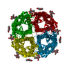 1j4nS S: Starting model for refinement |
|---|---|
| Similar structure data |

-Links
 PDBj
PDBj
- Assembly
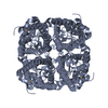
| Deposited unit | 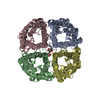
| ||||||||
|---|---|---|---|---|---|---|---|---|---|
| 1 |
| ||||||||
| Unit cell |
|
-Components
| #1: Protein | Mass: 28399.145 Da / Num. of mol.: 4 Source method: isolated from a genetically manipulated source Source: (gene. exp.) Homo sapiens (human) / Description: EasySelect Pichia Expression Kit (Invitrogen) / Gene: AQP5 / Plasmid: pPICZB / Production host:  Pichia pastoris (fungus) / Strain (production host): X33 / References: UniProt: P55064 Pichia pastoris (fungus) / Strain (production host): X33 / References: UniProt: P55064#2: Chemical | ChemComp-PS6 / |   Mass: 567.650 Da / Num. of mol.: 1 / Source method: obtained synthetically / Formula: C26H50NO10P Mass: 567.650 Da / Num. of mol.: 1 / Source method: obtained synthetically / Formula: C26H50NO10P#3: Water | ChemComp-HOH / |  Mass: 18.015 Da / Num. of mol.: 111 / Source method: isolated from a natural source / Formula: H2O Mass: 18.015 Da / Num. of mol.: 111 / Source method: isolated from a natural source / Formula: H2ONonpolymer details | PS6 IS A MODIFIED FORM OF PHOSPHATID | |
|---|
-Experimental details
-Experiment
| Experiment | Method: X-RAY DIFFRACTION / Number of used crystals: 1 |
|---|
- Sample preparation
| Crystal | Density Matthews: 3.15 Å3/Da / Density % sol: 61 % |
|---|---|
| Crystal grow | Temperature: 281 K / Method: vapor diffusion, hanging drop / pH: 7.6 Details: 19% polyethylene glycol 400, 0.1M Tris HCl pH 7.6, 0.1M NaCl, 6% v/v 1,6-hexanediol, 3% v/v 1,4-butanediol or 3% v/v 1,3-propanediol, VAPOR DIFFUSION, HANGING DROP, temperature 281K |
-Data collection
| Diffraction | Mean temperature: 170 K |
|---|---|
| Diffraction source | Source: SYNCHROTRON / Site: ESRF  / Beamline: ID14-3 / Wavelength: 0.931 Å / Beamline: ID14-3 / Wavelength: 0.931 Å |
| Detector | Type: ADSC QUANTUM 4 / Detector: CCD / Date: Jul 2, 2007 |
| Radiation | Protocol: SINGLE WAVELENGTH / Monochromatic (M) / Laue (L): M / Scattering type: x-ray |
| Radiation wavelength | Wavelength: 0.931 Å / Relative weight: 1 |
| Reflection | Resolution: 2→52.56 Å / Num. all: 84751 / Num. obs: 84751 / % possible obs: 82.9 % / Observed criterion σ(I): 3 / Redundancy: 3.7 % / Biso Wilson estimate: 26.15 Å2 / Rsym value: 0.061 / Net I/σ(I): 8.3 |
| Reflection shell | Resolution: 2→2.11 Å / Redundancy: 3.7 % / Mean I/σ(I) obs: 1.5 / Num. unique all: 12232 / Rsym value: 0.494 / % possible all: 82.6 |
- Processing
| Software |
| |||||||||||||||||||||||||||||||||
|---|---|---|---|---|---|---|---|---|---|---|---|---|---|---|---|---|---|---|---|---|---|---|---|---|---|---|---|---|---|---|---|---|---|---|
| Refinement | Method to determine structure: AB INITIO Starting model: PDB ENTRY 1J4N Resolution: 2→10 Å / Num. parameters: 29577 / Num. restraintsaints: 30558 / Cross valid method: FREE R / σ(F): 4 / Stereochemistry target values: Engh & Huber Details: Firstly, a homology model for AQP5 was generated using the SWISS-MODEL web server with the bovine AQP1 structure (PDB 1J4N) as a template. The homology model was then used as search model in ...Details: Firstly, a homology model for AQP5 was generated using the SWISS-MODEL web server with the bovine AQP1 structure (PDB 1J4N) as a template. The homology model was then used as search model in molecular replacement calculations. See primary citation for additional details.; This data is merohedrally twinned; THE TWINNING OPERATOR IS (H,K,L) -> (k, h, -l) AND THE TWINNING FRACTION IS 0.463. Diffraction data was from a crystal with near-perfect pseudo-merohedral twinning with the twin operator(010 100 00-1)swapping the a and b axes. Therefore, conventional programs/maps are not recommended when analysing this PDB and structure factors. ShelX (with BASF and TWIN values set) is useful for map calculation but the twinning compatible scripts are best for reliable omit maps (see Supporting Information of primary citation).
| |||||||||||||||||||||||||||||||||
| Displacement parameters | Biso mean: 37.2 Å2 | |||||||||||||||||||||||||||||||||
| Refine analyze | Num. disordered residues: 0 / Occupancy sum hydrogen: 0 / Occupancy sum non hydrogen: 7386.58 | |||||||||||||||||||||||||||||||||
| Refinement step | Cycle: LAST / Resolution: 2→10 Å
| |||||||||||||||||||||||||||||||||
| Refine LS restraints |
| |||||||||||||||||||||||||||||||||
| LS refinement shell | Resolution: 2→2.11 Å / Rfactor Rfree: 0.193 / Rfactor Rwork: 0.162 |
