 Movie
Movie Controller


+ Open data

- Basic information
| Entry | Database: PDB / ID: 4n82 | ||||||
|---|---|---|---|---|---|---|---|
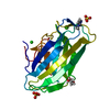
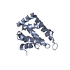
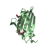
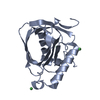
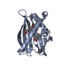
| Title | X-ray crystal structure of Streptococcus sanguinis NrdIox | ||||||
 Components Components | Ribonucleotide reductase | ||||||
 Keywords Keywords | OXIDOREDUCTASE / FLAVOPROTEIN / RIBONUCLEOTIDE REDUCTASE / OXIDATION-REDUCTION / FLAVIN MONONUCLEOTIDE | ||||||
| Function / homology |  Function and homology information Function and homology informationoxidoreductase activity, acting on NAD(P)H / protein modification process / FMN binding Similarity search - Function | ||||||
| Biological species |  Streptococcus sanguinis (bacteria) Streptococcus sanguinis (bacteria) | ||||||
| Method |  X-RAY DIFFRACTION / SYNCHROTRON / MOLECULAR REPLACEMENT / molecular replacement / Resolution: 1.88 Å X-RAY DIFFRACTION / SYNCHROTRON / MOLECULAR REPLACEMENT / molecular replacement / Resolution: 1.88 Å | ||||||
 Authors Authors | Boal, A.K. / Rosenzweig, A.C. | ||||||
 Citation Citation | Journal: J.Biol.Chem. / Year: 2014 Title: Streptococcus sanguinis Class Ib Ribonucleotide Reductase: HIGH ACTIVITY WITH BOTH IRON AND MANGANESE COFACTORS AND STRUCTURAL INSIGHTS. Authors: Makhlynets, O. / Boal, A.K. / Rhodes, D.V. / Kitten, T. / Rosenzweig, A.C. / Stubbe, J. | ||||||
| History |
|
- Structure visualization
| Structure viewer | Molecule: MolmilJmol/JSmol |
|---|
- Downloads & links
-Download
| PDBx/mmCIF format | 4n82.cif.gz | 138.9 KB | Display | PDBx/mmCIF format |
|---|---|---|---|---|
| PDB format | pdb4n82.ent.gz | 108.8 KB | Display | PDB format |
| PDBx/mmJSON format | 4n82.json.gz | Tree view | PDBx/mmJSON format | |
| Others |  Other downloads Other downloads |
-Validation report
| Summary document | 4n82_validation.pdf.gz | 1.1 MB | Display | wwPDB validaton report |
|---|---|---|---|---|
| Full document | 4n82_full_validation.pdf.gz | 1.1 MB | Display | |
| Data in XML | 4n82_validation.xml.gz | 15.7 KB | Display | |
| Data in CIF | 4n82_validation.cif.gz | 21.7 KB | Display | |
| Arichive directory | https://data.pdbj.org/pub/pdb/validation_reports/n8/4n82ftp://data.pdbj.org/pub/pdb/validation_reports/n8/4n82 | HTTPS FTP |
-Related structure data
| Related structure data |  4n83C 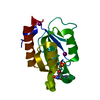 1rljS C: citing same article ( S: Starting model for refinement |
|---|---|
| Similar structure data |


-Links
 PDBj
PDBj


- Assembly
| Deposited unit | 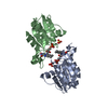
| ||||||||
|---|---|---|---|---|---|---|---|---|---|
| 1 | 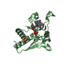
| ||||||||
| 2 | 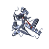
| ||||||||
| Unit cell |
|
-Components
| #1: Protein | Mass: 20069.586 Da / Num. of mol.: 2 Source method: isolated from a genetically manipulated source Source: (gene. exp.) Streptococcus sanguinis (bacteria) / Strain: SK36 / Gene: nrdI, SSA_2263 / Plasmid: pET28a / Production host: #2: Chemical |   Mass: 456.344 Da / Num. of mol.: 2 / Source method: obtained synthetically / Formula: C17H21N4O9P Mass: 456.344 Da / Num. of mol.: 2 / Source method: obtained synthetically / Formula: C17H21N4O9P#3: Chemical |   Mass: 96.063 Da / Num. of mol.: 2 / Source method: obtained synthetically / Formula: SO4 Mass: 96.063 Da / Num. of mol.: 2 / Source method: obtained synthetically / Formula: SO4#4: Water | ChemComp-HOH / |  Mass: 18.015 Da / Num. of mol.: 204 / Source method: isolated from a natural source / Formula: H2O Mass: 18.015 Da / Num. of mol.: 204 / Source method: isolated from a natural source / Formula: H2O |
|---|
-Experimental details
-Experiment
| Experiment | Method: X-RAY DIFFRACTION / Number of used crystals: 1 |
|---|
- Sample preparation
| Crystal | Density Matthews: 2.6 Å3/Da / Density % sol: 52.75 % |
|---|---|
| Crystal grow | Temperature: 298 K / Method: vapor diffusion, sitting drop / pH: 5.6 Details: PEG 4000, ammonium sulfate, pH 5.6, vapor diffusion, sitting drop, temperature 298K |
-Data collection
| Diffraction | Mean temperature: 100 K |
|---|---|
| Diffraction source | Source: SYNCHROTRON / Site: APS  / Beamline: 21-ID-D / Wavelength: 1.078 Å / Beamline: 21-ID-D / Wavelength: 1.078 Å |
| Detector | Type: MARRESEARCH / Detector: CCD / Date: Aug 19, 2012 |
| Radiation | Protocol: SINGLE WAVELENGTH / Monochromatic (M) / Laue (L): M / Scattering type: x-ray |
| Radiation wavelength | Wavelength: 1.078 Å / Relative weight: 1 |
| Reflection | Resolution: 1.88→50 Å / Num. obs: 33395 |
-Phasing
| Phasing | Method: molecular replacement |
|---|
- Processing
| Software |
| |||||||||||||||||||||||||||||||||||||||||||||||||||||||||||||||||||||||||||
|---|---|---|---|---|---|---|---|---|---|---|---|---|---|---|---|---|---|---|---|---|---|---|---|---|---|---|---|---|---|---|---|---|---|---|---|---|---|---|---|---|---|---|---|---|---|---|---|---|---|---|---|---|---|---|---|---|---|---|---|---|---|---|---|---|---|---|---|---|---|---|---|---|---|---|---|---|
| Refinement | Method to determine structure: MOLECULAR REPLACEMENT Starting model: PDB ENTRY 1RLJ Resolution: 1.88→47.69 Å / Cor.coef. Fo:Fc: 0.952 / Cor.coef. Fo:Fc free: 0.938 / Occupancy max: 1 / Occupancy min: 0.5 / SU B: 5.68 / SU ML: 0.094 / Cross valid method: THROUGHOUT / σ(F): 0 / ESU R: 0.143 / ESU R Free: 0.133 / Stereochemistry target values: MAXIMUM LIKELIHOOD / Details: HYDROGENS HAVE BEEN USED IF PRESENT IN THE INPUT
| |||||||||||||||||||||||||||||||||||||||||||||||||||||||||||||||||||||||||||
| Solvent computation | Ion probe radii: 0.8 Å / Shrinkage radii: 0.8 Å / VDW probe radii: 1.2 Å / Solvent model: MASK | |||||||||||||||||||||||||||||||||||||||||||||||||||||||||||||||||||||||||||
| Displacement parameters | Biso max: 74.85 Å2 / Biso mean: 33.6281 Å2 / Biso min: 16.87 Å2
| |||||||||||||||||||||||||||||||||||||||||||||||||||||||||||||||||||||||||||
| Refinement step | Cycle: LAST / Resolution: 1.88→47.69 Å
| |||||||||||||||||||||||||||||||||||||||||||||||||||||||||||||||||||||||||||
| Refine LS restraints |
| |||||||||||||||||||||||||||||||||||||||||||||||||||||||||||||||||||||||||||
| LS refinement shell | Resolution: 1.884→1.933 Å / Total num. of bins used: 20
| |||||||||||||||||||||||||||||||||||||||||||||||||||||||||||||||||||||||||||
| Refinement TLS params. | Method: refined / Refine-ID: X-RAY DIFFRACTION
| |||||||||||||||||||||||||||||||||||||||||||||||||||||||||||||||||||||||||||
| Refinement TLS group |
|
