 Movie
Movie Controller

[English] 日本語
 Yorodumi
- PDB-4n5b: Crystal structure of the Nipah virus phosphoprotein tetramerizati... -
+ Open data

- Basic information
| Entry | Database: PDB / ID: 4n5b | ||||||
|---|---|---|---|---|---|---|---|
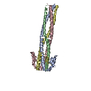
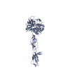
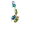
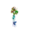
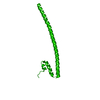
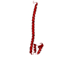
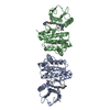
| Title | Crystal structure of the Nipah virus phosphoprotein tetramerization domain | ||||||
 Components Components | Phosphoprotein | ||||||
 Keywords Keywords | VIRAL PROTEIN / Viral Polymerase Co-Factor / Nucleoprotein Chaperone / Interferon Inhibitor | ||||||
| Function / homology |  Function and homology information Function and homology informationPhosphoprotein P region PNT disordered / Phosphoprotein P region PNT disordered / Phosphoprotein P soyouz module / N-terminal region of Paramyxovirinae phosphoprotein (P) / Paramyxovirus structural protein P/V, N-terminal domain / Paramyxovirus structural protein V/P N-terminus / P/V phosphoprotein, paramyxoviral / Paramyxovirus P/V phosphoprotein C-terminal Similarity search - Domain/homology | ||||||
| Biological species |  Nipah virus Nipah virus | ||||||
| Method |  X-RAY DIFFRACTION / SYNCHROTRON / AB INITIO PHASING / Resolution: 2.2 Å X-RAY DIFFRACTION / SYNCHROTRON / AB INITIO PHASING / Resolution: 2.2 Å | ||||||
 Authors Authors | Bruhn, J.F. / Barnett, K. / Bibby, J. / Thomas, J. / Keegan, R. / Rigden, D. / Bornholdt, Z.A. / Saphire, E.O. | ||||||
 Citation Citation | Journal: J.Virol. / Year: 2014 Title: Crystal structure of the nipah virus phosphoprotein tetramerization domain. Authors: Bruhn, J.F. / Barnett, K.C. / Bibby, J. / Thomas, J.M. / Keegan, R.M. / Rigden, D.J. / Bornholdt, Z.A. / Saphire, E.O. | ||||||
| History |
|
- Structure visualization
| Structure viewer | Molecule: MolmilJmol/JSmol |
|---|
- Downloads & links
-Download
| PDBx/mmCIF format | 4n5b.cif.gz | 192.1 KB | Display | PDBx/mmCIF format |
|---|---|---|---|---|
| PDB format | pdb4n5b.ent.gz | 153.9 KB | Display | PDB format |
| PDBx/mmJSON format | 4n5b.json.gz | Tree view | PDBx/mmJSON format | |
| Others |  Other downloads Other downloads |
-Validation report
| Arichive directory | https://data.pdbj.org/pub/pdb/validation_reports/n5/4n5bftp://data.pdbj.org/pub/pdb/validation_reports/n5/4n5b | HTTPS FTP |
|---|
-Related structure data
| Related structure data |  4eijS S: Starting model for refinement |
|---|---|
| Similar structure data |

-Links
 PDBj
PDBj


- Assembly
| Deposited unit | 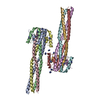
| ||||||||
|---|---|---|---|---|---|---|---|---|---|
| 1 | 
| ||||||||
| 2 | 
| ||||||||
| Unit cell |
| ||||||||
| Details | Chains A, B, C and D / Chains E, F, G and H |
-Components
| #1: Protein | Mass: 12622.337 Da / Num. of mol.: 8 / Fragment: Tetramerization Domain residues 470-578 Source method: isolated from a genetically manipulated source Source: (gene. exp.) Nipah virus / Strain: Ind-Nipah-07-FG / Gene: P/V/W/C / Plasmid: pET46 / Production host:  #2: Chemical | ChemComp-IMD /   Mass: 69.085 Da / Num. of mol.: 21 / Source method: obtained synthetically / Formula: C3H5N2 Mass: 69.085 Da / Num. of mol.: 21 / Source method: obtained synthetically / Formula: C3H5N2#3: Water | ChemComp-HOH / |  Mass: 18.015 Da / Num. of mol.: 860 / Source method: isolated from a natural source / Formula: H2O Mass: 18.015 Da / Num. of mol.: 860 / Source method: isolated from a natural source / Formula: H2O |
|---|
-Experimental details
-Experiment
| Experiment | Method: X-RAY DIFFRACTION / Number of used crystals: 1 |
|---|
- Sample preparation
| Crystal | Density Matthews: 2.67 Å3/Da / Density % sol: 53.93 % |
|---|---|
| Crystal grow | Temperature: 295.5 K / Method: vapor diffusion, sitting drop / pH: 7 Details: 0.1M imidazole pH 7.0, 25% PEG MME 550, 15% glycerol, VAPOR DIFFUSION, SITTING DROP, temperature 100K, temperature 295.5K |
-Data collection
| Diffraction | Mean temperature: 100 K |
|---|---|
| Diffraction source | Source: SYNCHROTRON / Site: ALS  / Beamline: 5.0.2 / Wavelength: 1 Å / Beamline: 5.0.2 / Wavelength: 1 Å |
| Detector | Type: ADSC QUANTUM 315r / Detector: CCD / Date: Jun 9, 2013 |
| Radiation | Monochromator: Double-crystal, Si(111) liquid N2 cooled / Protocol: SINGLE WAVELENGTH / Monochromatic (M) / Laue (L): M / Scattering type: x-ray |
| Radiation wavelength | Wavelength: 1 Å / Relative weight: 1 |
| Reflection | Resolution: 2.2→46.37 Å / Num. obs: 51813 / % possible obs: 97.7 % / Observed criterion σ(F): 1 / Observed criterion σ(I): 1 / Rmerge(I) obs: 0.063 / Net I/σ(I): 9.3 |
| Reflection shell | Resolution: 2.2→2.28 Å / Redundancy: 3.85 % / Rmerge(I) obs: 0.298 / Mean I/σ(I) obs: 3 / % possible all: 97.2 |
- Processing
| Software |
| ||||||||||||||||||||||||||||||||||||||||||||||||||||||||||||||||||||||||||||||||||||||||||||||||||||||||||||||||||||||||||||||||||||||||||||
|---|---|---|---|---|---|---|---|---|---|---|---|---|---|---|---|---|---|---|---|---|---|---|---|---|---|---|---|---|---|---|---|---|---|---|---|---|---|---|---|---|---|---|---|---|---|---|---|---|---|---|---|---|---|---|---|---|---|---|---|---|---|---|---|---|---|---|---|---|---|---|---|---|---|---|---|---|---|---|---|---|---|---|---|---|---|---|---|---|---|---|---|---|---|---|---|---|---|---|---|---|---|---|---|---|---|---|---|---|---|---|---|---|---|---|---|---|---|---|---|---|---|---|---|---|---|---|---|---|---|---|---|---|---|---|---|---|---|---|---|---|---|
| Refinement | Method to determine structure: AB INITIO PHASING Starting model: PDB ENTRY 4EIJ Resolution: 2.2→46.369 Å / SU ML: 0.23 / σ(F): 1.97 / Phase error: 27.07 / Stereochemistry target values: ML
| ||||||||||||||||||||||||||||||||||||||||||||||||||||||||||||||||||||||||||||||||||||||||||||||||||||||||||||||||||||||||||||||||||||||||||||
| Solvent computation | Shrinkage radii: 0.9 Å / VDW probe radii: 1.11 Å / Solvent model: FLAT BULK SOLVENT MODEL | ||||||||||||||||||||||||||||||||||||||||||||||||||||||||||||||||||||||||||||||||||||||||||||||||||||||||||||||||||||||||||||||||||||||||||||
| Refinement step | Cycle: LAST / Resolution: 2.2→46.369 Å
| ||||||||||||||||||||||||||||||||||||||||||||||||||||||||||||||||||||||||||||||||||||||||||||||||||||||||||||||||||||||||||||||||||||||||||||
| Refine LS restraints |
| ||||||||||||||||||||||||||||||||||||||||||||||||||||||||||||||||||||||||||||||||||||||||||||||||||||||||||||||||||||||||||||||||||||||||||||
| LS refinement shell |
|
