 Movie
Movie Controller

[English] 日本語
 Yorodumi
- PDB-1zjk: Crystal structure of the zymogen catalytic region of human MASP-2 -
+ Open data

- Basic information
| Entry | Database: PDB / ID: 1zjk | ||||||
|---|---|---|---|---|---|---|---|
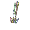
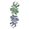
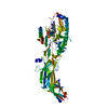
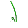
| Title | Crystal structure of the zymogen catalytic region of human MASP-2 | ||||||
 Components Components | Mannan-binding lectin serine protease 2 | ||||||
 Keywords Keywords | HYDROLASE / beta barrel / modular protein | ||||||
| Function / homology |  Function and homology information Function and homology informationmannan-binding lectin-associated serine protease-2 / complement component C4b binding / Ficolins bind to repetitive carbohydrate structures on the target cell surface / Lectin pathway of complement activation / complement activation, lectin pathway / Initial triggering of complement / complement activation, classical pathway / calcium-dependent protein binding / peptidase activity / serine-type endopeptidase activity ...mannan-binding lectin-associated serine protease-2 / complement component C4b binding / Ficolins bind to repetitive carbohydrate structures on the target cell surface / Lectin pathway of complement activation / complement activation, lectin pathway / Initial triggering of complement / complement activation, classical pathway / calcium-dependent protein binding / peptidase activity / serine-type endopeptidase activity / calcium ion binding / SARS-CoV-2 activates/modulates innate and adaptive immune responses / proteolysis / : / extracellular exosome / extracellular region / identical protein binding Similarity search - Function | ||||||
| Biological species |  Homo sapiens (human) Homo sapiens (human) | ||||||
| Method |  X-RAY DIFFRACTION / SYNCHROTRON / MOLECULAR REPLACEMENT / Resolution: 2.18 Å X-RAY DIFFRACTION / SYNCHROTRON / MOLECULAR REPLACEMENT / Resolution: 2.18 Å | ||||||
 Authors Authors | Gal, P. / Harmat, V. / Kocsis, A. / Bian, T. / Barna, L. / Ambrus, G. / Vegh, B. / Balczer, J. / Sim, R.B. / Naray-Szabo, G. / Zavodszky, P. | ||||||
 Citation Citation | Journal: J.Biol.Chem. / Year: 2005 Title: A True Autoactivating Enzyme: Structural insight into mannose-binding lectin-associated serine protease-2 activations Authors: Gal, P. / Harmat, V. / Kocsis, A. / Bian, T. / Barna, L. / Ambrus, G. / Vegh, B. / Balczer, J. / Sim, R.B. / Naray-Szabo, G. / Zavodszky, P. #1: Journal: J.Mol.Biol. / Year: 2004Title: The Structure of MBL-Associated Serine Protease-2 Reveals that Identical Substrate Specificities of C1s and MASP-2 are Realized Through Different Sets of Enzyme-Substrate Interactions Authors: Harmat, V. / Gal, P. / Kardos, J. / Szilagyi, K. / Ambrus, G. / Vegh, B. / Naray-Szabo, G. / Zavodszky, P. | ||||||
| History |
|
- Structure visualization
| Structure viewer | Molecule: MolmilJmol/JSmol |
|---|
- Downloads & links
-Download
| PDBx/mmCIF format | 1zjk.cif.gz | 89.2 KB | Display | PDBx/mmCIF format |
|---|---|---|---|---|
| PDB format | pdb1zjk.ent.gz | 66.5 KB | Display | PDB format |
| PDBx/mmJSON format | 1zjk.json.gz | Tree view | PDBx/mmJSON format | |
| Others |  Other downloads Other downloads |
-Validation report
| Arichive directory | https://data.pdbj.org/pub/pdb/validation_reports/zj/1zjkftp://data.pdbj.org/pub/pdb/validation_reports/zj/1zjk | HTTPS FTP |
|---|
-Related structure data
| Related structure data |  1q3xS S: Starting model for refinement |
|---|---|
| Similar structure data |


-Links
 PDBj
PDBj


- Assembly
| Deposited unit | 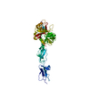
| ||||||||
|---|---|---|---|---|---|---|---|---|---|
| 1 |
| ||||||||
| Unit cell |
|
-Components
| #1: Protein | Mass: 44031.715 Da / Num. of mol.: 1 Fragment: complement control protein modules 1,2 and serine protease domain Mutation: R444Q Source method: isolated from a genetically manipulated source Source: (gene. exp.) Homo sapiens (human) / Gene: MASP2 / Plasmid: pET-17b / Species (production host): Escherichia coli / Production host:  References: UniProt: O00187, Hydrolases; Acting on peptide bonds (peptidases); Serine endopeptidases |
|---|---|
| #2: Water | ChemComp-HOH /  Mass: 18.015 Da / Num. of mol.: 84 / Source method: isolated from a natural source / Formula: H2O Mass: 18.015 Da / Num. of mol.: 84 / Source method: isolated from a natural source / Formula: H2O |
| Has protein modification | Y |
-Experimental details
-Experiment
| Experiment | Method: X-RAY DIFFRACTION / Number of used crystals: 1 |
|---|
- Sample preparation
| Crystal | Density Matthews: 2.2 Å3/Da / Density % sol: 43.3 % |
|---|---|
| Crystal grow | Temperature: 293 K / Method: vapor diffusion, hanging drop / pH: 8 Details: PEG 6000, sodium chloride, TRIS/HCl buffer, pH 8.0, temperature 293K, VAPOR DIFFUSION, HANGING DROP |
-Data collection
| Diffraction | Mean temperature: 100 K |
|---|---|
| Diffraction source | Source: SYNCHROTRON / Site: ESRF  / Beamline: ID14-4 / Wavelength: 0.9392 Å / Beamline: ID14-4 / Wavelength: 0.9392 Å |
| Detector | Type: ADSC QUANTUM 4 / Detector: CCD / Date: Mar 18, 2003 / Details: toroidal focusing mirror |
| Radiation | Monochromator: double crystal monochromator / Protocol: SINGLE WAVELENGTH / Monochromatic (M) / Laue (L): M / Scattering type: x-ray |
| Radiation wavelength | Wavelength: 0.9392 Å / Relative weight: 1 |
| Reflection | Resolution: 2.18→28.9 Å / Num. all: 18330 / Num. obs: 18330 / % possible obs: 88.1 % / Observed criterion σ(I): -3 / Redundancy: 8.3 % / Rmerge(I) obs: 0.099 / Net I/σ(I): 15.13 |
| Reflection shell | Resolution: 2.18→2.25 Å / Redundancy: 1.9 % / Rmerge(I) obs: 0.575 / Mean I/σ(I) obs: 1.9 / Num. unique all: 800 / % possible all: 43.1 |
- Processing
| Software |
| ||||||||||||||||||||||||||||||||||||||||||||||||||||||||||||||||||||||||||||||||||||||||||||||||||||
|---|---|---|---|---|---|---|---|---|---|---|---|---|---|---|---|---|---|---|---|---|---|---|---|---|---|---|---|---|---|---|---|---|---|---|---|---|---|---|---|---|---|---|---|---|---|---|---|---|---|---|---|---|---|---|---|---|---|---|---|---|---|---|---|---|---|---|---|---|---|---|---|---|---|---|---|---|---|---|---|---|---|---|---|---|---|---|---|---|---|---|---|---|---|---|---|---|---|---|---|---|---|
| Refinement | Method to determine structure: MOLECULAR REPLACEMENT Starting model: SP domain of PDB ENTRY 1Q3X Resolution: 2.18→28.9 Å / Cor.coef. Fo:Fc: 0.942 / Cor.coef. Fo:Fc free: 0.908 / SU B: 8.011 / SU ML: 0.194 / TLS residual ADP flag: LIKELY RESIDUAL / Cross valid method: THROUGHOUT / σ(F): 0 / ESU R: 0.352 / ESU R Free: 0.243 / Stereochemistry target values: MAXIMUM LIKELIHOOD / Details: HYDROGENS HAVE BEEN ADDED IN THE RIDING POSITIONS
| ||||||||||||||||||||||||||||||||||||||||||||||||||||||||||||||||||||||||||||||||||||||||||||||||||||
| Solvent computation | Ion probe radii: 0.8 Å / Shrinkage radii: 0.8 Å / VDW probe radii: 1.4 Å / Solvent model: BABINET MODEL WITH MASK | ||||||||||||||||||||||||||||||||||||||||||||||||||||||||||||||||||||||||||||||||||||||||||||||||||||
| Displacement parameters | Biso mean: 25.915 Å2
| ||||||||||||||||||||||||||||||||||||||||||||||||||||||||||||||||||||||||||||||||||||||||||||||||||||
| Refinement step | Cycle: LAST / Resolution: 2.18→28.9 Å
| ||||||||||||||||||||||||||||||||||||||||||||||||||||||||||||||||||||||||||||||||||||||||||||||||||||
| Refine LS restraints |
| ||||||||||||||||||||||||||||||||||||||||||||||||||||||||||||||||||||||||||||||||||||||||||||||||||||
| LS refinement shell | Resolution: 2.18→2.237 Å / Total num. of bins used: 20 /
| ||||||||||||||||||||||||||||||||||||||||||||||||||||||||||||||||||||||||||||||||||||||||||||||||||||
| Refinement TLS params. | Method: refined / Refine-ID: X-RAY DIFFRACTION
| ||||||||||||||||||||||||||||||||||||||||||||||||||||||||||||||||||||||||||||||||||||||||||||||||||||
| Refinement TLS group |
|
