 Movie
Movie Controller

[English] 日本語
 Yorodumi
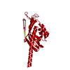
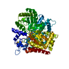
- PDB-4n3v: Crystal structure of a bile-acid 7-alpha dehydratase (CLOHIR_0007... -
+ Open data

- Basic information
| Entry | Database: PDB / ID: 4n3v | ||||||
|---|---|---|---|---|---|---|---|
| Title | Crystal structure of a bile-acid 7-alpha dehydratase (CLOHIR_00079) from Clostridium hiranonis DSM 13275 at 1.89 A resolution with product added | ||||||
 Components Components | Bile acid 7-alpha dehydratase, BaiE | ||||||
 Keywords Keywords | LYASE / SnoaL-like domain / PF13577 family / Structural Genomics / Joint Center for Structural Genomics / JCSG / Protein Structure Initiative / PSI-BIOLOGY | ||||||
| Function / homology |  Function and homology information Function and homology informationbile-acid 7alpha-dehydratase / bile-acid 7alpha-dehydratase activity / metal ion binding Similarity search - Function | ||||||
| Biological species |  Clostridium hiranonis (bacteria) Clostridium hiranonis (bacteria) | ||||||
| Method |  X-RAY DIFFRACTION / SYNCHROTRON / MOLECULAR REPLACEMENT / molecular replacement / Resolution: 1.89 Å X-RAY DIFFRACTION / SYNCHROTRON / MOLECULAR REPLACEMENT / molecular replacement / Resolution: 1.89 Å | ||||||
 Authors Authors | Joint Center for Structural Genomics (JCSG) | ||||||
 Citation Citation | Journal: To be published Title: Crystal structure of a bile-acid 7-alpha dehydratase (CLOHIR_00079) from Clostridium hiranonis DSM 13275 at 1.89 A resolution with product added Authors: Joint Center for Structural Genomics (JCSG) | ||||||
| History |
|
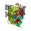
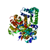
- Structure visualization
| Structure viewer | Molecule: MolmilJmol/JSmol |
|---|
- Downloads & links
-Download
| PDBx/mmCIF format | 4n3v.cif.gz | 163.6 KB | Display | PDBx/mmCIF format |
|---|---|---|---|---|
| PDB format | pdb4n3v.ent.gz | 128.5 KB | Display | PDB format |
| PDBx/mmJSON format | 4n3v.json.gz | Tree view | PDBx/mmJSON format | |
| Others |  Other downloads Other downloads |
-Validation report
| Arichive directory | https://data.pdbj.org/pub/pdb/validation_reports/n3/4n3vftp://data.pdbj.org/pub/pdb/validation_reports/n3/4n3v | HTTPS FTP |
|---|
-Related structure data
| Related structure data | 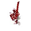 4l8pS S: Starting model for refinement |
|---|---|
| Similar structure data | |
| Other databases |


-Links
 PDBj
PDBj


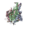
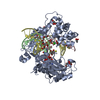
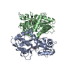
- Assembly
| Deposited unit | 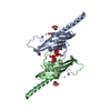
| |||||||||||||||||||||||||||||||||||||||
|---|---|---|---|---|---|---|---|---|---|---|---|---|---|---|---|---|---|---|---|---|---|---|---|---|---|---|---|---|---|---|---|---|---|---|---|---|---|---|---|---|
| 1 | 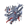
| |||||||||||||||||||||||||||||||||||||||
| 2 | 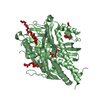
| |||||||||||||||||||||||||||||||||||||||
| Unit cell |
| |||||||||||||||||||||||||||||||||||||||
| Components on special symmetry positions |
| |||||||||||||||||||||||||||||||||||||||
| Noncrystallographic symmetry (NCS) | NCS domain:
NCS domain segments: Component-ID: _ / Ens-ID: 1 / Beg auth comp-ID: GLY / Beg label comp-ID: GLY / End auth comp-ID: UNL / End label comp-ID: UNL / Refine code: _ / Auth seq-ID: 0 - 180 / Label seq-ID: 19
|
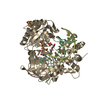
-Components
-Protein , 1 types, 2 molecules AB
| #1: Protein | Mass: 21958.766 Da / Num. of mol.: 2 Source method: isolated from a genetically manipulated source Source: (gene. exp.) Clostridium hiranonis (bacteria) / Strain: DSM 13275 / Gene: baiE, CLOHIR_00079 / Plasmid: SpeedET / Production host: |
|---|
-Non-polymers , 7 types, 199 molecules 


| #2: Chemical |  Mass: 65.409 Da / Num. of mol.: 2 / Source method: obtained synthetically / Formula: Zn Mass: 65.409 Da / Num. of mol.: 2 / Source method: obtained synthetically / Formula: Zn#3: Chemical |  Mass: 22.990 Da / Num. of mol.: 2 / Source method: obtained synthetically / Formula: Na Mass: 22.990 Da / Num. of mol.: 2 / Source method: obtained synthetically / Formula: Na#4: Chemical |  Mass: 192.124 Da / Num. of mol.: 2 / Source method: obtained synthetically / Formula: C6H8O7 Mass: 192.124 Da / Num. of mol.: 2 / Source method: obtained synthetically / Formula: C6H8O7#5: Chemical | ChemComp-EDO / |  Mass: 62.068 Da / Num. of mol.: 1 / Source method: obtained synthetically / Formula: C2H6O2 Mass: 62.068 Da / Num. of mol.: 1 / Source method: obtained synthetically / Formula: C2H6O2#6: Chemical | ChemComp-1PE / |  Mass: 238.278 Da / Num. of mol.: 1 / Source method: obtained synthetically / Formula: C10H22O6 / Comment: precipitant*YM Mass: 238.278 Da / Num. of mol.: 1 / Source method: obtained synthetically / Formula: C10H22O6 / Comment: precipitant*YM#7: Chemical | Num. of mol.: 2 / Source method: obtained synthetically #8: Water | ChemComp-HOH / | Mass: 18.015 Da / Num. of mol.: 189 / Source method: isolated from a natural source / Formula: H2O |
|---|
-Details
| Sequence details | THIS CONSTRUCT (RESIDUES 1-168) WAS EXPRESSED WITH AN N-TERMINAL PURIFICATI |
|---|
-Experimental details
-Experiment
| Experiment | Method: X-RAY DIFFRACTION / Number of used crystals: 1 |
|---|
- Sample preparation
| Crystal | Density Matthews: 2.43 Å3/Da / Density % sol: 49.31 % |
|---|---|
| Crystal grow | Temperature: 293 K / Method: vapor diffusion, sitting drop / pH: 5 Details: 10.00% polyethylene glycol 6000, 0.1M citric acid pH 5.0, Additive: 0.001 M 3-oxo-delta 4,6, Lithocholyl Coenzyme A, NANODROP', VAPOR DIFFUSION, SITTING DROP, temperature 293K |
-Data collection
| Diffraction | Mean temperature: 100 K | |||||||||||||||||||||||||||||||||||||||||||||||||||||||||||||||||||||||||||||||||||||||||||||||||||||||||||||||||||||||||||||||||||||||||||||||||||||||||||||||||||||||||||||||||||||||||||||
|---|---|---|---|---|---|---|---|---|---|---|---|---|---|---|---|---|---|---|---|---|---|---|---|---|---|---|---|---|---|---|---|---|---|---|---|---|---|---|---|---|---|---|---|---|---|---|---|---|---|---|---|---|---|---|---|---|---|---|---|---|---|---|---|---|---|---|---|---|---|---|---|---|---|---|---|---|---|---|---|---|---|---|---|---|---|---|---|---|---|---|---|---|---|---|---|---|---|---|---|---|---|---|---|---|---|---|---|---|---|---|---|---|---|---|---|---|---|---|---|---|---|---|---|---|---|---|---|---|---|---|---|---|---|---|---|---|---|---|---|---|---|---|---|---|---|---|---|---|---|---|---|---|---|---|---|---|---|---|---|---|---|---|---|---|---|---|---|---|---|---|---|---|---|---|---|---|---|---|---|---|---|---|---|---|---|---|---|---|---|---|
| Diffraction source | Source: SYNCHROTRON / Site: ALS  / Beamline: 8.2.2 / Wavelength: 0.999947 / Beamline: 8.2.2 / Wavelength: 0.999947 | |||||||||||||||||||||||||||||||||||||||||||||||||||||||||||||||||||||||||||||||||||||||||||||||||||||||||||||||||||||||||||||||||||||||||||||||||||||||||||||||||||||||||||||||||||||||||||||
| Detector | Type: ADSC QUANTUM 315 / Detector: CCD / Date: Sep 19, 2012 / Details: KOHZU: Double Crystal Si(111) | |||||||||||||||||||||||||||||||||||||||||||||||||||||||||||||||||||||||||||||||||||||||||||||||||||||||||||||||||||||||||||||||||||||||||||||||||||||||||||||||||||||||||||||||||||||||||||||
| Radiation | Monochromator: Double Crystal Si(111) / Protocol: SINGLE WAVELENGTH / Monochromatic (M) / Laue (L): M / Scattering type: x-ray | |||||||||||||||||||||||||||||||||||||||||||||||||||||||||||||||||||||||||||||||||||||||||||||||||||||||||||||||||||||||||||||||||||||||||||||||||||||||||||||||||||||||||||||||||||||||||||||
| Radiation wavelength | Wavelength: 0.999947 Å / Relative weight: 1 | |||||||||||||||||||||||||||||||||||||||||||||||||||||||||||||||||||||||||||||||||||||||||||||||||||||||||||||||||||||||||||||||||||||||||||||||||||||||||||||||||||||||||||||||||||||||||||||
| Reflection | Resolution: 1.89→47.414 Å / Num. obs: 34715 / % possible obs: 99.9 % / Observed criterion σ(I): -3 / Biso Wilson estimate: 26.185 Å2 / Rmerge F obs: 0.999 / Rmerge(I) obs: 0.069 / Rrim(I) all: 0.073 / Net I/σ(I): 19.5 / Num. measured all: 362385 | |||||||||||||||||||||||||||||||||||||||||||||||||||||||||||||||||||||||||||||||||||||||||||||||||||||||||||||||||||||||||||||||||||||||||||||||||||||||||||||||||||||||||||||||||||||||||||||
| Reflection shell | Diffraction-ID: 1
|
-Phasing
| Phasing | Method: molecular replacement |
|---|
- Processing
| Software |
| |||||||||||||||||||||||||||||||||||||||||||||||||||||||||||||||||||||||||||
|---|---|---|---|---|---|---|---|---|---|---|---|---|---|---|---|---|---|---|---|---|---|---|---|---|---|---|---|---|---|---|---|---|---|---|---|---|---|---|---|---|---|---|---|---|---|---|---|---|---|---|---|---|---|---|---|---|---|---|---|---|---|---|---|---|---|---|---|---|---|---|---|---|---|---|---|---|
| Refinement | Method to determine structure: MOLECULAR REPLACEMENT Starting model: 4L8P Resolution: 1.89→47.414 Å / Cor.coef. Fo:Fc: 0.963 / Cor.coef. Fo:Fc free: 0.95 / Occupancy max: 1 / Occupancy min: 0.33 / SU B: 5.543 / SU ML: 0.086 / Cross valid method: THROUGHOUT / σ(F): 0 / ESU R: 0.141 / ESU R Free: 0.129 / Stereochemistry target values: MAXIMUM LIKELIHOOD Details: 1. HYDROGENS HAVE BEEN ADDED IN THE RIDING POSITIONS. 2. ATOM RECORDS CONTAIN SUM OF TLS AND RESIDUAL B FACTORS. 3. ANISOU RECORDS CONTAIN SUM OF TLS AND RESIDUAL U FACTORS. 4. WATERS WERE ...Details: 1. HYDROGENS HAVE BEEN ADDED IN THE RIDING POSITIONS. 2. ATOM RECORDS CONTAIN SUM OF TLS AND RESIDUAL B FACTORS. 3. ANISOU RECORDS CONTAIN SUM OF TLS AND RESIDUAL U FACTORS. 4. WATERS WERE EXCLUDED FROM AUTOMATIC TLS ASSIGNMENT. 5. AN UNKNOWN LIGAND (UNL) HAS BEEN MODELED IN A POCKET FORMED BY PROTOMERS FROM TWO ADJACENT TRIMERS IN THE CRYSTAL LATTICE. THIS UNL MAY REPRESENT THE ADDED PRODUCT COMPOUND, 3-OXO-DELTA 4,6, LITHOCHOLYL COENZYME A. HOWEVER, THE 2-FOLD DISORDER OF THE SITE AND LIKELY PARTIAL OCCUPANCY PREVENTS DEFANITIVIE ASSIGNEMNT OF THESE DENSITY FEATURES. 6. A ZINC ION (ZN2+) IS MODELED BASED ON A PEAK IN THE ANOMALOUS DIFFERENCE FOURIER MAP. THE ZINC ASSIGNMENT WAS TENATIVLY BASED ON THE PRESENCE OF ENDOGENOUS ZINC IN A HOMOLOG STRUCTURE AND COULD REPRESENT A DIFFERENT METAL ION. X-RAY FLUORESCENCE SPECTRA WERE NOT AVAILABLE FOR THIS CRYSTAL. 7. THE MODELED CITRIC ACID (CIT), PEG FRAGMENT (1PE), 1,2- ETHANEDIOL AND SODIUM ION (NA1+) ARE PRESENT IN THE CRYSTALLIZATION SOLUTION, CRYO SOLUTION AND PROTEIN BUFFER. 8. THE SCATTERING FACTORS FOR SODIUM, SULFUR, AND ZINC ATOMS WERE ADJUSTED BY REFMAC 5.7.0032 TO ACCOUNT FOR ANOMALOUS DISPERSION BASED ON THE WAVELENGTH 0.9999 A (NA F'= 0.06, S F'= 0.19, ZN F'= -0.35). THE CROMER MANN VALUES LISTED IN THE CIF VERSION OF THE FILE INCLUDE THIS CORRECTION.
| |||||||||||||||||||||||||||||||||||||||||||||||||||||||||||||||||||||||||||
| Solvent computation | Ion probe radii: 0.8 Å / Shrinkage radii: 0.8 Å / VDW probe radii: 1.2 Å / Solvent model: BABINET MODEL WITH MASK | |||||||||||||||||||||||||||||||||||||||||||||||||||||||||||||||||||||||||||
| Displacement parameters | Biso max: 108.78 Å2 / Biso mean: 35.823 Å2 / Biso min: 15.11 Å2
| |||||||||||||||||||||||||||||||||||||||||||||||||||||||||||||||||||||||||||
| Refinement step | Cycle: LAST / Resolution: 1.89→47.414 Å
| |||||||||||||||||||||||||||||||||||||||||||||||||||||||||||||||||||||||||||
| Refine LS restraints |
| |||||||||||||||||||||||||||||||||||||||||||||||||||||||||||||||||||||||||||
| Refine LS restraints NCS | Ens-ID: 1 / Number: 24935 / Refine-ID: X-RAY DIFFRACTION / Type: interatomic distance / Rms dev position: 0.03 Å / Weight position: 0.05
| |||||||||||||||||||||||||||||||||||||||||||||||||||||||||||||||||||||||||||
| LS refinement shell | Resolution: 1.89→1.939 Å / Total num. of bins used: 20
| |||||||||||||||||||||||||||||||||||||||||||||||||||||||||||||||||||||||||||
| Refinement TLS params. | Method: refined / Refine-ID: X-RAY DIFFRACTION
| |||||||||||||||||||||||||||||||||||||||||||||||||||||||||||||||||||||||||||
| Refinement TLS group |
|
