 Movie
Movie Controller


+ Open data

- Basic information
| Entry | Database: PDB / ID: 3mt5 | ||||||
|---|---|---|---|---|---|---|---|
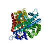
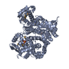
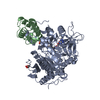
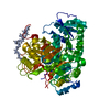
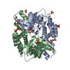
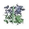
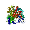
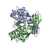
| Title | Crystal Structure of the Human BK Gating Apparatus | ||||||
 Components Components | Potassium large conductance calcium-activated channel, subfamily M, alpha member 1 | ||||||
 Keywords Keywords | MEMBRANE PROTEIN / TRANSPORT PROTEIN / potassium channel | ||||||
| Function / homology |  Function and homology information Function and homology informationmicturition / Acetylcholine inhibits contraction of outer hair cells / large conductance calcium-activated potassium channel activity / Ca2+ activated K+ channels / calcium-activated potassium channel activity / negative regulation of cell volume / smooth muscle contraction involved in micturition / response to carbon monoxide / response to osmotic stress / Sensory processing of sound by inner hair cells of the cochlea ...micturition / Acetylcholine inhibits contraction of outer hair cells / large conductance calcium-activated potassium channel activity / Ca2+ activated K+ channels / calcium-activated potassium channel activity / negative regulation of cell volume / smooth muscle contraction involved in micturition / response to carbon monoxide / response to osmotic stress / Sensory processing of sound by inner hair cells of the cochlea / cGMP effects / intracellular potassium ion homeostasis / voltage-gated potassium channel activity / voltage-gated potassium channel complex / potassium ion transmembrane transport / potassium ion transport / regulation of membrane potential / response to calcium ion / caveola / vasodilation / actin binding / response to hypoxia / postsynaptic membrane / apical plasma membrane / positive regulation of apoptotic process / membrane / metal ion binding / identical protein binding / plasma membrane Similarity search - Function | ||||||
| Biological species |  Homo sapiens (human) Homo sapiens (human) | ||||||
| Method |  X-RAY DIFFRACTION / SYNCHROTRON / MAD / Resolution: 3 Å X-RAY DIFFRACTION / SYNCHROTRON / MAD / Resolution: 3 Å | ||||||
 Authors Authors | Yuan, P. / MacKinnon, R. | ||||||
 Citation Citation | Journal: Science / Year: 2010 Title: Structure of the human BK channel Ca2+-activation apparatus at 3.0 A resolution. Authors: Yuan, P. / Leonetti, M.D. / Pico, A.R. / Hsiung, Y. / MacKinnon, R. | ||||||
| History |
|
- Structure visualization
| Structure viewer | Molecule: MolmilJmol/JSmol |
|---|
- Downloads & links
-Download
| PDBx/mmCIF format | 3mt5.cif.gz | 132.3 KB | Display | PDBx/mmCIF format |
|---|---|---|---|---|
| PDB format | pdb3mt5.ent.gz | 99.4 KB | Display | PDB format |
| PDBx/mmJSON format | 3mt5.json.gz | Tree view | PDBx/mmJSON format | |
| Others |  Other downloads Other downloads |
-Validation report
| Arichive directory | https://data.pdbj.org/pub/pdb/validation_reports/mt/3mt5ftp://data.pdbj.org/pub/pdb/validation_reports/mt/3mt5 | HTTPS FTP |
|---|
-Related structure data
| Similar structure data |
|---|


-Links
 PDBj
PDBj


- Assembly
| Deposited unit | 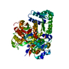
| ||||||||
|---|---|---|---|---|---|---|---|---|---|
| 1 |
| ||||||||
| 2 | x 6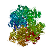
| ||||||||
| Unit cell |
|
-Components
| #1: Protein | Mass: 81644.406 Da / Num. of mol.: 1 / Fragment: Cytoplasmic Domain (UNP RESIDUED 356-1071) Source method: isolated from a genetically manipulated source Source: (gene. exp.) Homo sapiens (human) / Gene: KCNMA1, RP11-443A13.1-004 / Plasmid: pFastBac / Production host:   Spodoptera frugiperda (fall armyworm) / Strain (production host): sf9 / References: UniProt: Q5SVK2, UniProt: Q12791*PLUS Spodoptera frugiperda (fall armyworm) / Strain (production host): sf9 / References: UniProt: Q5SVK2, UniProt: Q12791*PLUS |
|---|---|
| #2: Chemical | ChemComp-CA /   Mass: 40.078 Da / Num. of mol.: 1 / Source method: obtained synthetically / Formula: Ca Mass: 40.078 Da / Num. of mol.: 1 / Source method: obtained synthetically / Formula: Ca |
| #3: Chemical | ChemComp-SO4 /   Mass: 96.063 Da / Num. of mol.: 1 / Source method: obtained synthetically / Formula: SO4 Mass: 96.063 Da / Num. of mol.: 1 / Source method: obtained synthetically / Formula: SO4 |
-Experimental details
-Experiment
| Experiment | Method: X-RAY DIFFRACTION / Number of used crystals: 1 |
|---|
- Sample preparation
| Crystal | Density Matthews: 3.36 Å3/Da / Density % sol: 63.43 % |
|---|---|
| Crystal grow | Temperature: 293 K / Method: vapor diffusion, hanging drop / pH: 4.8 Details: PEG 4000, pH 4.8, VAPOR DIFFUSION, HANGING DROP, temperature 293K |
-Data collection
| Diffraction |
| |||||||||||||||||||||||||||||||||||||||||||||||||||||||||||||||||||||||||||||
|---|---|---|---|---|---|---|---|---|---|---|---|---|---|---|---|---|---|---|---|---|---|---|---|---|---|---|---|---|---|---|---|---|---|---|---|---|---|---|---|---|---|---|---|---|---|---|---|---|---|---|---|---|---|---|---|---|---|---|---|---|---|---|---|---|---|---|---|---|---|---|---|---|---|---|---|---|---|---|
| Diffraction source |
| |||||||||||||||||||||||||||||||||||||||||||||||||||||||||||||||||||||||||||||
| Detector |
| |||||||||||||||||||||||||||||||||||||||||||||||||||||||||||||||||||||||||||||
| Radiation |
| |||||||||||||||||||||||||||||||||||||||||||||||||||||||||||||||||||||||||||||
| Radiation wavelength |
| |||||||||||||||||||||||||||||||||||||||||||||||||||||||||||||||||||||||||||||
| Reflection | Redundancy: 7 % / Av σ(I) over netI: 19.6 / Number: 122548 / Rmerge(I) obs: 0.105 / Χ2: 1.39 / D res high: 3.3 Å / D res low: 50 Å / Num. obs: 17611 / % possible obs: 99.8 | |||||||||||||||||||||||||||||||||||||||||||||||||||||||||||||||||||||||||||||
| Diffraction reflection shell |
| |||||||||||||||||||||||||||||||||||||||||||||||||||||||||||||||||||||||||||||
| Reflection | Resolution: 3→50 Å / Num. obs: 23149 / % possible obs: 99.9 % / Redundancy: 5.8 % / Rmerge(I) obs: 0.089 / Χ2: 1.077 / Net I/σ(I): 9.4 | |||||||||||||||||||||||||||||||||||||||||||||||||||||||||||||||||||||||||||||
| Reflection shell |
|

-Phasing
| Phasing | Method: MAD | ||||||||||||||||||||||||||||||||||||||||||||||||||||||||||||||||||||||||||||||||||||||||||||||||||
|---|---|---|---|---|---|---|---|---|---|---|---|---|---|---|---|---|---|---|---|---|---|---|---|---|---|---|---|---|---|---|---|---|---|---|---|---|---|---|---|---|---|---|---|---|---|---|---|---|---|---|---|---|---|---|---|---|---|---|---|---|---|---|---|---|---|---|---|---|---|---|---|---|---|---|---|---|---|---|---|---|---|---|---|---|---|---|---|---|---|---|---|---|---|---|---|---|---|---|---|
| Phasing set |
| ||||||||||||||||||||||||||||||||||||||||||||||||||||||||||||||||||||||||||||||||||||||||||||||||||
| Phasing MAD set |
| ||||||||||||||||||||||||||||||||||||||||||||||||||||||||||||||||||||||||||||||||||||||||||||||||||
| Phasing MAD set site |
| ||||||||||||||||||||||||||||||||||||||||||||||||||||||||||||||||||||||||||||||||||||||||||||||||||
| Phasing dm | FOM : 0.7 / FOM acentric: 0.7 / FOM centric: 0.72 / Reflection: 16652 / Reflection acentric: 14118 / Reflection centric: 2534 | ||||||||||||||||||||||||||||||||||||||||||||||||||||||||||||||||||||||||||||||||||||||||||||||||||
| Phasing dm shell |
|
- Processing
| Software |
| |||||||||||||||||||||||||||||||||||||||||||||||||||||||||||||||||
|---|---|---|---|---|---|---|---|---|---|---|---|---|---|---|---|---|---|---|---|---|---|---|---|---|---|---|---|---|---|---|---|---|---|---|---|---|---|---|---|---|---|---|---|---|---|---|---|---|---|---|---|---|---|---|---|---|---|---|---|---|---|---|---|---|---|---|
| Refinement | Method to determine structure: MAD / Resolution: 3→47.5 Å / Cor.coef. Fo:Fc: 0.908 / Cor.coef. Fo:Fc free: 0.898 / WRfactor Rfree: 0.265 / WRfactor Rwork: 0.234 / Occupancy max: 1 / Occupancy min: 1 / FOM work R set: 0.825 / SU B: 17.116 / SU ML: 0.314 / SU R Cruickshank DPI: 0.91 / SU Rfree: 0.393 / Cross valid method: THROUGHOUT / σ(F): 0 / ESU R Free: 0.393 Stereochemistry target values: MAXIMUM LIKELIHOOD WITH PHASES Details: HYDROGENS HAVE BEEN ADDED IN THE RIDING POSITIONS U VALUES : REFINED INDIVIDUALLY
| |||||||||||||||||||||||||||||||||||||||||||||||||||||||||||||||||
| Solvent computation | Ion probe radii: 0.8 Å / Shrinkage radii: 0.8 Å / VDW probe radii: 1.4 Å / Solvent model: MASK | |||||||||||||||||||||||||||||||||||||||||||||||||||||||||||||||||
| Displacement parameters | Biso max: 120.18 Å2 / Biso mean: 67.341 Å2 / Biso min: 34.06 Å2
| |||||||||||||||||||||||||||||||||||||||||||||||||||||||||||||||||
| Refinement step | Cycle: LAST / Resolution: 3→47.5 Å
| |||||||||||||||||||||||||||||||||||||||||||||||||||||||||||||||||
| Refine LS restraints |
| |||||||||||||||||||||||||||||||||||||||||||||||||||||||||||||||||
| LS refinement shell | Resolution: 3→3.078 Å / Total num. of bins used: 20
|
