 Movie
Movie Controller

[English] 日本語
 Yorodumi
- PDB-4mzk: Crystal Structure of MTIP from Plasmodium falciparum in complex w... -
+ Open data

- Basic information
| Entry | Database: PDB / ID: 4mzk | ||||||
|---|---|---|---|---|---|---|---|
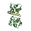
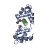
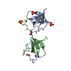
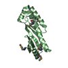
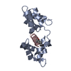
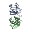
| Title | Crystal Structure of MTIP from Plasmodium falciparum in complex with pGly[807,811], a stapled myoA tail peptide | ||||||
 Components Components |
| ||||||
 Keywords Keywords | PROTEIN BINDING/inhibitor / Actomyosin motor / Stapled peptides / PROTEIN BINDING-inhibitor complex | ||||||
| Function / homology |  Function and homology information Function and homology informationpellicle / inner membrane pellicle complex / glideosome / myosin complex / myosin II complex / microfilament motor activity / cytoskeletal motor activity / actin filament organization / actin filament binding / actin cytoskeleton ...pellicle / inner membrane pellicle complex / glideosome / myosin complex / myosin II complex / microfilament motor activity / cytoskeletal motor activity / actin filament organization / actin filament binding / actin cytoskeleton / actin binding / calcium ion binding / ATP binding / membrane / plasma membrane / cytoplasm Similarity search - Function | ||||||
| Biological species |  | ||||||
| Method |  X-RAY DIFFRACTION / SYNCHROTRON / MOLECULAR REPLACEMENT / Resolution: 1.82 Å X-RAY DIFFRACTION / SYNCHROTRON / MOLECULAR REPLACEMENT / Resolution: 1.82 Å | ||||||
 Authors Authors | Douse, C.H. / Garnett, J.A. / Maas, S.J. / Cota, E. / Tate, E.W. | ||||||
 Citation Citation | Journal: Acs Chem.Biol. / Year: 2014 Title: Crystal Structures of Stapled and Hydrogen Bond Surrogate Peptides Targeting a Fully Buried Protein-Helix Interaction. Authors: Douse, C.H. / Maas, S.J. / Thomas, J.C. / Garnett, J.A. / Sun, Y. / Cota, E. / Tate, E.W. | ||||||
| History |
|
- Structure visualization
| Structure viewer | Molecule: MolmilJmol/JSmol |
|---|
- Downloads & links
-Download
| PDBx/mmCIF format | 4mzk.cif.gz | 81.1 KB | Display | PDBx/mmCIF format |
|---|---|---|---|---|
| PDB format | pdb4mzk.ent.gz | 60.1 KB | Display | PDB format |
| PDBx/mmJSON format | 4mzk.json.gz | Tree view | PDBx/mmJSON format | |
| Others |  Other downloads Other downloads |
-Validation report
| Arichive directory | https://data.pdbj.org/pub/pdb/validation_reports/mz/4mzkftp://data.pdbj.org/pub/pdb/validation_reports/mz/4mzk | HTTPS FTP |
|---|
-Related structure data
| Related structure data | 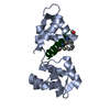 4mzjC  4mzlC 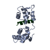 4aomS S: Starting model for refinement C: citing same article ( |
|---|---|
| Similar structure data |


-Links
 PDBj
PDBj


- Assembly
| Deposited unit | 
| ||||||||
|---|---|---|---|---|---|---|---|---|---|
| 1 |
| ||||||||
| Unit cell |
|
-Components
| #1: Protein | Mass: 16498.234 Da / Num. of mol.: 1 / Fragment: unp residues 61-204 Source method: isolated from a genetically manipulated source Source: (gene. exp.) Strain: 3D7 / Gene: MTIP, PFL2225w / Plasmid: pRSETA / Production host:  |
|---|---|
| #2: Protein/peptide |   Type: Peptide-like / Class: Inhibitor / Mass: 2177.744 Da / Num. of mol.: 1 / Fragment: unp residues 799-816 / Source method: obtained synthetically Type: Peptide-like / Class: Inhibitor / Mass: 2177.744 Da / Num. of mol.: 1 / Fragment: unp residues 799-816 / Source method: obtained syntheticallyDetails: Synthesised peptide mimic of Plasmodium falciparum myosin A tail using Fmoc-Solid Phase Peptide Synthesis Source: (synth.) References: UniProt: Q8IDR3, pGly[807,811], a stapled myoA tail peptide |
| #3: Water | ChemComp-HOH /  Mass: 18.015 Da / Num. of mol.: 101 / Source method: isolated from a natural source / Formula: H2O Mass: 18.015 Da / Num. of mol.: 101 / Source method: isolated from a natural source / Formula: H2O |
| Has protein modification | Y |
| Sequence details | THE PEPTIDE CORRESPONDING TO CHAIN T IS A STAPLED PEPTIDE. THE NLE T 807 AND NLE T 811 ARE CYCLIZED ...THE PEPTIDE CORRESPOND |
-Experimental details
-Experiment
| Experiment | Method: X-RAY DIFFRACTION / Number of used crystals: 1 |
|---|
- Sample preparation
| Crystal | Density Matthews: 2.12 Å3/Da / Density % sol: 42.02 % |
|---|---|
| Crystal grow | Temperature: 293 K / Method: vapor diffusion, sitting drop / pH: 6.5 Details: Reservoir solution: 200 mM ammonium acetate, 10 mM magnesium acetate, 50 mM sodium cacodylate trihydrate, 30 % PEG 8000, pH 6.5, VAPOR DIFFUSION, SITTING DROP, temperature 293K |
-Data collection
| Diffraction | Mean temperature: 100 K | |||||||||||||||||||||
|---|---|---|---|---|---|---|---|---|---|---|---|---|---|---|---|---|---|---|---|---|---|---|
| Diffraction source | Source: SYNCHROTRON / Site: Diamond  / Beamline: I02 / Wavelength: 0.979 Å / Beamline: I02 / Wavelength: 0.979 Å | |||||||||||||||||||||
| Detector | Type: PSI PILATUS 6M / Detector: PIXEL / Date: Apr 25, 2013 | |||||||||||||||||||||
| Radiation | Monochromator: Double crystal Si(111) / Protocol: SINGLE WAVELENGTH / Monochromatic (M) / Laue (L): M / Scattering type: x-ray | |||||||||||||||||||||
| Radiation wavelength | Wavelength: 0.979 Å / Relative weight: 1 | |||||||||||||||||||||
| Reflection | Resolution: 1.82→37.98 Å / Num. all: 14866 / Num. obs: 14865 / % possible obs: 100 % / Observed criterion σ(I): 2 / Redundancy: 12.9 % / Rmerge(I) obs: 0.069 / Net I/σ(I): 26.2 | |||||||||||||||||||||
| Reflection shell |
|
- Processing
| Software |
| |||||||||||||||||||||||||||||||||||||||||||||||||||||||||||||||||||||||||||||||||||||||||||||||||||||||||||||||||||||||||||||||||||||||||||||||||||||||||||||||||||||||||||||||||||||||||||||||||||||||||||||||||||||||||||||||||||||||||||||||||||||||||||||||||||||||||||||||||||||||||||||||||||||||||||||||||||||||||||||||||||||
|---|---|---|---|---|---|---|---|---|---|---|---|---|---|---|---|---|---|---|---|---|---|---|---|---|---|---|---|---|---|---|---|---|---|---|---|---|---|---|---|---|---|---|---|---|---|---|---|---|---|---|---|---|---|---|---|---|---|---|---|---|---|---|---|---|---|---|---|---|---|---|---|---|---|---|---|---|---|---|---|---|---|---|---|---|---|---|---|---|---|---|---|---|---|---|---|---|---|---|---|---|---|---|---|---|---|---|---|---|---|---|---|---|---|---|---|---|---|---|---|---|---|---|---|---|---|---|---|---|---|---|---|---|---|---|---|---|---|---|---|---|---|---|---|---|---|---|---|---|---|---|---|---|---|---|---|---|---|---|---|---|---|---|---|---|---|---|---|---|---|---|---|---|---|---|---|---|---|---|---|---|---|---|---|---|---|---|---|---|---|---|---|---|---|---|---|---|---|---|---|---|---|---|---|---|---|---|---|---|---|---|---|---|---|---|---|---|---|---|---|---|---|---|---|---|---|---|---|---|---|---|---|---|---|---|---|---|---|---|---|---|---|---|---|---|---|---|---|---|---|---|---|---|---|---|---|---|---|---|---|---|---|---|---|---|---|---|---|---|---|---|---|---|---|---|---|---|---|---|---|---|---|---|---|---|---|---|---|---|---|---|---|---|---|---|---|---|---|---|---|---|---|---|---|---|---|---|---|---|---|---|---|---|---|---|---|---|---|---|---|---|---|---|---|---|---|---|
| Refinement | Method to determine structure: MOLECULAR REPLACEMENT Starting model: pdb entry 4AOM Resolution: 1.82→37.915 Å / SU ML: 0.26 / σ(F): 1.49 / Phase error: 21.78 / Stereochemistry target values: ML
| |||||||||||||||||||||||||||||||||||||||||||||||||||||||||||||||||||||||||||||||||||||||||||||||||||||||||||||||||||||||||||||||||||||||||||||||||||||||||||||||||||||||||||||||||||||||||||||||||||||||||||||||||||||||||||||||||||||||||||||||||||||||||||||||||||||||||||||||||||||||||||||||||||||||||||||||||||||||||||||||||||||
| Solvent computation | Shrinkage radii: 0.9 Å / VDW probe radii: 1.11 Å / Solvent model: FLAT BULK SOLVENT MODEL / Bsol: 27.279 Å2 / ksol: 0.31 e/Å3 | |||||||||||||||||||||||||||||||||||||||||||||||||||||||||||||||||||||||||||||||||||||||||||||||||||||||||||||||||||||||||||||||||||||||||||||||||||||||||||||||||||||||||||||||||||||||||||||||||||||||||||||||||||||||||||||||||||||||||||||||||||||||||||||||||||||||||||||||||||||||||||||||||||||||||||||||||||||||||||||||||||||
| Refinement step | Cycle: LAST / Resolution: 1.82→37.915 Å
| |||||||||||||||||||||||||||||||||||||||||||||||||||||||||||||||||||||||||||||||||||||||||||||||||||||||||||||||||||||||||||||||||||||||||||||||||||||||||||||||||||||||||||||||||||||||||||||||||||||||||||||||||||||||||||||||||||||||||||||||||||||||||||||||||||||||||||||||||||||||||||||||||||||||||||||||||||||||||||||||||||||
| Refine LS restraints |
| |||||||||||||||||||||||||||||||||||||||||||||||||||||||||||||||||||||||||||||||||||||||||||||||||||||||||||||||||||||||||||||||||||||||||||||||||||||||||||||||||||||||||||||||||||||||||||||||||||||||||||||||||||||||||||||||||||||||||||||||||||||||||||||||||||||||||||||||||||||||||||||||||||||||||||||||||||||||||||||||||||||
| LS refinement shell |
| |||||||||||||||||||||||||||||||||||||||||||||||||||||||||||||||||||||||||||||||||||||||||||||||||||||||||||||||||||||||||||||||||||||||||||||||||||||||||||||||||||||||||||||||||||||||||||||||||||||||||||||||||||||||||||||||||||||||||||||||||||||||||||||||||||||||||||||||||||||||||||||||||||||||||||||||||||||||||||||||||||||
| Refinement TLS params. | Method: refined / Refine-ID: X-RAY DIFFRACTION
| |||||||||||||||||||||||||||||||||||||||||||||||||||||||||||||||||||||||||||||||||||||||||||||||||||||||||||||||||||||||||||||||||||||||||||||||||||||||||||||||||||||||||||||||||||||||||||||||||||||||||||||||||||||||||||||||||||||||||||||||||||||||||||||||||||||||||||||||||||||||||||||||||||||||||||||||||||||||||||||||||||||
| Refinement TLS group |
|
