Monochromator: double crystal / Protocol: SINGLE WAVELENGTH / Monochromatic (M) / Laue (L): M / Scattering type: x-ray
Radiation wavelength
Wavelength: 0.97915 Å / Relative weight: 1
Reflection
Resolution: 1.35→30 Å / Num. all: 76260 / Num. obs: 76134 / % possible obs: 99.8 % / Observed criterion σ(I): -3 / Redundancy: 7.1 % / Biso Wilson estimate: 15.4 Å2 / Rmerge(I) obs: 0.084 / Net I/σ(I): 26.8
Reflection shell
Resolution: 1.35→1.37 Å / Redundancy: 6.9 % / Rmerge(I) obs: 0.891 / Mean I/σ(I) obs: 2 / Num. unique all: 3769 / % possible all: 99.9
-
Processing
Software
Name
Version
Classification
SBC-Collect
datacollection
SHELX
modelbuilding
MLPHARE
phasing
DM
modelbuilding
ARP/wARP
modelbuilding
Coot
modelbuilding
PHENIX
(phenix.refine: dev_1446)
refinement
HKL-3000
datareduction
HKL-3000
datascaling
SHELX
phasing
DM
phasing
Refinement
Method to determine structure: SAD / Resolution: 1.349→24.898 Å / SU ML: 0.12 / Isotropic thermal model: anisotropic / Cross valid method: THROUGHOUT / σ(F): 0 / Phase error: 15.92 / Stereochemistry target values: ML / Details: hydrogen atoms have been added at riding positions
Rfactor
Num. reflection
% reflection
Selection details
Rfree
0.1589
1113
1.46 %
random
Rwork
0.1355
-
-
-
all
0.1359
76091
-
-
obs
0.1359
76091
99.74 %
-
Solvent computation
Shrinkage radii: 0.9 Å / VDW probe radii: 1.11 Å / Solvent model: FLAT BULK SOLVENT MODEL
Refinement step
Cycle: LAST / Resolution: 1.349→24.898 Å
Protein
Nucleic acid
Ligand
Solvent
Total
Num. atoms
2028
0
3
289
2320
Refine LS restraints
Refine-ID
Type
Dev ideal
Number
X-RAY DIFFRACTION
f_bond_d
0.013
2429
X-RAY DIFFRACTION
f_angle_d
1.461
3335
X-RAY DIFFRACTION
f_dihedral_angle_d
13.313
893
X-RAY DIFFRACTION
f_chiral_restr
0.095
349
X-RAY DIFFRACTION
f_plane_restr
0.007
444
LS refinement shell
Resolution (Å)
Rfactor Rfree
Num. reflection Rfree
Rfactor Rwork
Num. reflection Rwork
Refine-ID
% reflection obs (%)
1.3493-1.4107
0.2369
125
0.2017
9240
X-RAY DIFFRACTION
99
1.4107-1.485
0.1896
155
0.1516
9274
X-RAY DIFFRACTION
100
1.485-1.578
0.1728
136
0.1227
9330
X-RAY DIFFRACTION
100
1.578-1.6999
0.1192
123
0.103
9330
X-RAY DIFFRACTION
100
1.6999-1.8709
0.1443
144
0.1003
9347
X-RAY DIFFRACTION
100
1.8709-2.1415
0.1481
127
0.109
9423
X-RAY DIFFRACTION
100
2.1415-2.6975
0.1448
154
0.1353
9417
X-RAY DIFFRACTION
100
2.6975-24.9019
0.1685
149
0.1544
9617
X-RAY DIFFRACTION
99
+
About Yorodumi
-
News
-
Feb 9, 2022. New format data for meta-information of EMDB entries
New format data for meta-information of EMDB entries
Version 3 of the EMDB header file is now the official format.
The previous official version 1.9 will be removed from the archive.
In the structure databanks used in Yorodumi, some data are registered as the other names, "COVID-19 virus" and "2019-nCoV". Here are the details of the virus and the list of structure data.
Jan 31, 2019. EMDB accession codes are about to change! (news from PDBe EMDB page)
EMDB accession codes are about to change! (news from PDBe EMDB page)
The allocation of 4 digits for EMDB accession codes will soon come to an end. Whilst these codes will remain in use, new EMDB accession codes will include an additional digit and will expand incrementally as the available range of codes is exhausted. The current 4-digit format prefixed with “EMD-” (i.e. EMD-XXXX) will advance to a 5-digit format (i.e. EMD-XXXXX), and so on. It is currently estimated that the 4-digit codes will be depleted around Spring 2019, at which point the 5-digit format will come into force.
The EM Navigator/Yorodumi systems omit the EMD- prefix.
Related info.:Q: What is EMD? / ID/Accession-code notation in Yorodumi/EM Navigator
Yorodumi is a browser for structure data from EMDB, PDB, SASBDB, etc.
This page is also the successor to EM Navigator detail page, and also detail information page/front-end page for Omokage search.
The word "yorodu" (or yorozu) is an old Japanese word meaning "ten thousand". "mi" (miru) is to see.
Related info.:EMDB / PDB / SASBDB / Comparison of 3 databanks / Yorodumi Search / Aug 31, 2016. New EM Navigator & Yorodumi / Yorodumi Papers / Jmol/JSmol / Function and homology information / Changes in new EM Navigator and Yorodumi
 Movie
Movie Controller

 Yorodumi
 Open data
 Basic information
 Components
 Keywords
 Function and homology information
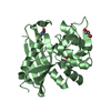
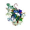
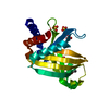
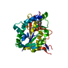
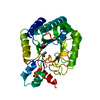
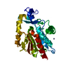
 Photorhabdus luminescens subsp. laumondii (bacteria)
 X-RAY DIFFRACTION /
  Authors
 Citation
 Structure visualization
 Downloads & links
 Other downloads
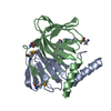


 PDBj
PDBj Assembly

 Mass: 58.693 Da / Num. of mol.: 2 / Source method: obtained synthetically / Formula: Ni
 Mass: 22.990 Da / Num. of mol.: 1 / Source method: obtained synthetically / Formula: Na
 Mass: 18.015 Da / Num. of mol.: 289 / Source method: isolated from a natural source / Formula: H2O
 Sample preparation
 / Beamline: 19-ID / Wavelength: 0.97915 Å
 Processing