 Movie
Movie Controller

[English] 日本語
 Yorodumi
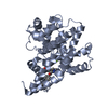
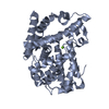
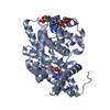
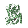
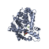
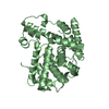
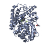
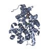
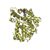
- PDB-4msh: Crystal Structure of PDE10A2 with fragment ZT0143 ((2S)-4-chloro-... -
+ Open data

- Basic information
| Entry | Database: PDB / ID: 4msh | ||||||
|---|---|---|---|---|---|---|---|
| Title | Crystal Structure of PDE10A2 with fragment ZT0143 ((2S)-4-chloro-2,3-dihydro-1,3-benzothiazol-2-amine) | ||||||
 Components Components | cAMP and cAMP-inhibited cGMP 3',5'-cyclic phosphodiesterase 10A | ||||||
 Keywords Keywords | HYDROLASE/HYDROLASE INHIBITOR / fragment screening / hydrolase / HYDROLASE-HYDROLASE INHIBITOR complex | ||||||
| Function / homology |  Function and homology information Function and homology information3',5'-cGMP-stimulated cyclic-nucleotide phosphodiesterase activity / 3',5'-cyclic-nucleotide phosphodiesterase / negative regulation of receptor guanylyl cyclase signaling pathway / cGMP catabolic process / cAMP catabolic process / cGMP effects / 3',5'-cyclic-nucleotide phosphodiesterase activity / cGMP binding / 3',5'-cyclic-GMP phosphodiesterase activity / 3',5'-cyclic-AMP phosphodiesterase activity ...3',5'-cGMP-stimulated cyclic-nucleotide phosphodiesterase activity / 3',5'-cyclic-nucleotide phosphodiesterase / negative regulation of receptor guanylyl cyclase signaling pathway / cGMP catabolic process / cAMP catabolic process / cGMP effects / 3',5'-cyclic-nucleotide phosphodiesterase activity / cGMP binding / 3',5'-cyclic-GMP phosphodiesterase activity / 3',5'-cyclic-AMP phosphodiesterase activity / regulation of adenylate cyclase-activating G protein-coupled receptor signaling pathway / cAMP binding / negative regulation of cAMP/PKA signal transduction / G alpha (s) signalling events / glutamatergic synapse / signal transduction / metal ion binding / cytosol Similarity search - Function | ||||||
| Biological species |  Homo sapiens (human) Homo sapiens (human) | ||||||
| Method |  X-RAY DIFFRACTION / MOLECULAR REPLACEMENT / Resolution: 2.3 Å X-RAY DIFFRACTION / MOLECULAR REPLACEMENT / Resolution: 2.3 Å | ||||||
 Authors Authors | Sridhar, V. / Badger, J. / Logan, C. / Chie-Leon, B. / Nienaber, V. | ||||||
 Citation Citation | Journal: J Biomol Screen / Year: 2014 Title: Identification and optimization of PDE10A inhibitors using fragment-based screening by nanocalorimetry and X-ray crystallography. Authors: Recht, M.I. / Sridhar, V. / Badger, J. / Bounaud, P.Y. / Logan, C. / Chie-Leon, B. / Nienaber, V. / Torres, F.E. | ||||||
| History |
|
- Structure visualization
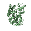
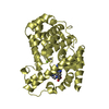
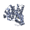
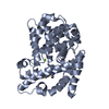
| Structure viewer | Molecule: MolmilJmol/JSmol |
|---|
- Downloads & links
-Download
| PDBx/mmCIF format | 4msh.cif.gz | 141.9 KB | Display | PDBx/mmCIF format |
|---|---|---|---|---|
| PDB format | pdb4msh.ent.gz | 111.2 KB | Display | PDB format |
| PDBx/mmJSON format | 4msh.json.gz | Tree view | PDBx/mmJSON format | |
| Others |  Other downloads Other downloads |
-Validation report
| Arichive directory | https://data.pdbj.org/pub/pdb/validation_reports/ms/4mshftp://data.pdbj.org/pub/pdb/validation_reports/ms/4msh | HTTPS FTP |
|---|
-Related structure data
| Related structure data |  4lkqC  4lljC  4llkC  4llpC  4llxC  4lm0C 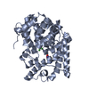 4lm1C 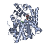 4lm2C 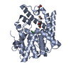 4lm3C  4lm4C 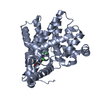 4mrwC  4mrzC  4ms0C  4msaC 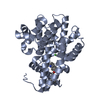 4mscC  4mseC 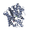 4msnC  2ourS C: citing same article ( S: Starting model for refinement |
|---|---|
| Similar structure data |

-Links
 PDBj
PDBj

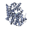
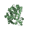
- Assembly
| Deposited unit | 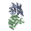
| ||||||||
|---|---|---|---|---|---|---|---|---|---|
| 1 | 
| ||||||||
| 2 | 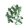
| ||||||||
| Unit cell |
|
-Components
| #1: Protein | Mass: 39479.242 Da / Num. of mol.: 2 / Fragment: catalytic domain, UNP residues 439-779 Source method: isolated from a genetically manipulated source Source: (gene. exp.) Homo sapiens (human) / Gene: PDE10A / Production host:  References: UniProt: Q9Y233, 3',5'-cyclic-nucleotide phosphodiesterase, 3',5'-cyclic-GMP phosphodiesterase #2: Chemical | ChemComp-2D0 / | 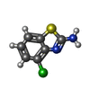  Mass: 184.646 Da / Num. of mol.: 1 / Source method: obtained synthetically / Formula: C7H5ClN2S Mass: 184.646 Da / Num. of mol.: 1 / Source method: obtained synthetically / Formula: C7H5ClN2S#3: Chemical | ChemComp-NI /   Mass: 58.693 Da / Num. of mol.: 4 / Source method: obtained synthetically / Formula: Ni Mass: 58.693 Da / Num. of mol.: 4 / Source method: obtained synthetically / Formula: Ni#4: Water | ChemComp-HOH / |  Mass: 18.015 Da / Num. of mol.: 16 / Source method: isolated from a natural source / Formula: H2O Mass: 18.015 Da / Num. of mol.: 16 / Source method: isolated from a natural source / Formula: H2ONonpolymer details | THE FOUR DIVALENT CATIONS ARE REPRESENTE | |
|---|
-Experimental details
-Experiment
| Experiment | Method: X-RAY DIFFRACTION / Number of used crystals: 1 |
|---|
- Sample preparation
| Crystal | Density Matthews: 2.03 Å3/Da / Density % sol: 39.45 % |
|---|---|
| Crystal grow | Temperature: 277 K / Method: vapor diffusion, hanging drop / pH: 7.5 Details: 18% PEG 4450, 0.2M calcium acetate, 50mM BME, pH 7.5, VAPOR DIFFUSION, HANGING DROP, temperature 277K |
-Data collection
| Diffraction | Mean temperature: 100 K |
|---|---|
| Diffraction source | Source: ROTATING ANODE / Type: RIGAKU MICROMAX-007 HF / Wavelength: 1.54 Å |
| Detector | Type: MAR scanner 345 mm plate / Detector: IMAGE PLATE / Date: Mar 10, 2011 |
| Radiation | Protocol: SINGLE WAVELENGTH / Monochromatic (M) / Laue (L): M / Scattering type: x-ray |
| Radiation wavelength | Wavelength: 1.54 Å / Relative weight: 1 |
| Reflection | Resolution: 2.3→39.69 Å / Num. all: 20880 / Num. obs: 20880 / % possible obs: 70.7 % / Observed criterion σ(F): -4 / Observed criterion σ(I): -4 / Redundancy: 5 % / Rmerge(I) obs: 0.131 / Net I/σ(I): 9 |
| Reflection shell | Resolution: 2.3→2.42 Å / Redundancy: 5.9 % / Rmerge(I) obs: 0.706 / Mean I/σ(I) obs: 2.3 / Num. unique all: 16120 / % possible all: 64.8 |
- Processing
| Software |
| |||||||||||||||||||||||||||||||||||||||||||||||||||||||||||||||||
|---|---|---|---|---|---|---|---|---|---|---|---|---|---|---|---|---|---|---|---|---|---|---|---|---|---|---|---|---|---|---|---|---|---|---|---|---|---|---|---|---|---|---|---|---|---|---|---|---|---|---|---|---|---|---|---|---|---|---|---|---|---|---|---|---|---|---|
| Refinement | Method to determine structure: MOLECULAR REPLACEMENT Starting model: PDB entry 2OUR Resolution: 2.3→39.69 Å / Cor.coef. Fo:Fc: 0.908 / Cor.coef. Fo:Fc free: 0.84 / SU B: 9.978 / SU ML: 0.245 / Cross valid method: THROUGHOUT / ESU R Free: 0.352 / Stereochemistry target values: MAXIMUM LIKELIHOOD
| |||||||||||||||||||||||||||||||||||||||||||||||||||||||||||||||||
| Solvent computation | Ion probe radii: 0.8 Å / Shrinkage radii: 0.8 Å / VDW probe radii: 1.4 Å / Solvent model: MASK | |||||||||||||||||||||||||||||||||||||||||||||||||||||||||||||||||
| Displacement parameters | Biso mean: 29.737 Å2
| |||||||||||||||||||||||||||||||||||||||||||||||||||||||||||||||||
| Refinement step | Cycle: LAST / Resolution: 2.3→39.69 Å
| |||||||||||||||||||||||||||||||||||||||||||||||||||||||||||||||||
| Refine LS restraints |
| |||||||||||||||||||||||||||||||||||||||||||||||||||||||||||||||||
| LS refinement shell | Resolution: 2.3→2.36 Å / Total num. of bins used: 20 /
|
