 Movie
Movie Controller

[English] 日本語
 Yorodumi
- PDB-4mjz: 2.75 Angstrom Resolution Crystal Structure of Putative Orotidine-... -
+ Open data

- Basic information
| Entry | Database: PDB / ID: 4mjz | ||||||
|---|---|---|---|---|---|---|---|
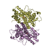
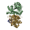
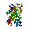
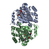
| Title | 2.75 Angstrom Resolution Crystal Structure of Putative Orotidine-monophosphate-decarboxylase from Toxoplasma gondii. | ||||||
 Components Components | Orotidine 5'-phosphate decarboxylase | ||||||
 Keywords Keywords | LYASE / Structural Genomics / NIAID / National Institute of Allergy and Infectious Diseases / Center for Structural Genomics of Infectious Diseases / CSGID | ||||||
| Function / homology |  Function and homology information Function and homology informationorotidine-5'-phosphate decarboxylase / orotidine-5'-phosphate decarboxylase activity / glycosyltransferase activity / 'de novo' UMP biosynthetic process / 'de novo' pyrimidine nucleobase biosynthetic process Similarity search - Function | ||||||
| Biological species |  | ||||||
| Method |  X-RAY DIFFRACTION / SYNCHROTRON / MOLECULAR REPLACEMENT / Resolution: 2.75 Å X-RAY DIFFRACTION / SYNCHROTRON / MOLECULAR REPLACEMENT / Resolution: 2.75 Å | ||||||
 Authors Authors | Minasov, G. / Wawrzak, Z. / Ruan, J. / Ngo, H. / Shuvalova, L. / Dubrovska, I. / Anderson, W.F. / Center for Structural Genomics of Infectious Diseases (CSGID) | ||||||
 Citation Citation | Journal: TO BE PUBLISHED Title: 2.75 Angstrom Resolution Crystal Structure of Putative Orotidine-monophosphate-decarboxylase from Toxoplasma gondii. Authors: Minasov, G. / Wawrzak, Z. / Ruan, J. / Ngo, H. / Shuvalova, L. / Dubrovska, I. / Anderson, W.F. / Center for Structural Genomics of Infectious Diseases (CSGID) | ||||||
| History |
|
- Structure visualization
| Structure viewer | Molecule: MolmilJmol/JSmol |
|---|
- Downloads & links
-Download
| PDBx/mmCIF format | 4mjz.cif.gz | 233.7 KB | Display | PDBx/mmCIF format |
|---|---|---|---|---|
| PDB format | pdb4mjz.ent.gz | 189.8 KB | Display | PDB format |
| PDBx/mmJSON format | 4mjz.json.gz | Tree view | PDBx/mmJSON format | |
| Others |  Other downloads Other downloads |
-Validation report
| Summary document | 4mjz_validation.pdf.gz | 473.5 KB | Display | wwPDB validaton report |
|---|---|---|---|---|
| Full document | 4mjz_full_validation.pdf.gz | 485 KB | Display | |
| Data in XML | 4mjz_validation.xml.gz | 23.6 KB | Display | |
| Data in CIF | 4mjz_validation.cif.gz | 32 KB | Display | |
| Arichive directory | https://data.pdbj.org/pub/pdb/validation_reports/mj/4mjzftp://data.pdbj.org/pub/pdb/validation_reports/mj/4mjz | HTTPS FTP |
-Related structure data
| Related structure data |  3qw4S S: Starting model for refinement |
|---|---|
| Similar structure data | |
| Other databases |
-Links
 PDBj
PDBj
- Assembly
| Deposited unit | 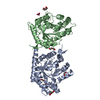
| ||||||||||||
|---|---|---|---|---|---|---|---|---|---|---|---|---|---|
| 1 | 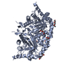
| ||||||||||||
| 2 | 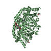
| ||||||||||||
| Unit cell |
| ||||||||||||
| Components on special symmetry positions |
| ||||||||||||
| Noncrystallographic symmetry (NCS) | NCS oper:
|
-Components
| #1: Protein | Mass: 35041.387 Da / Num. of mol.: 2 / Fragment: UNP residues 12-318 Source method: isolated from a genetically manipulated source Source: (gene. exp.)  References: UniProt: B9PKU1, UniProt: B6KBH9*PLUS, orotidine-5'-phosphate decarboxylase #2: Chemical | ChemComp-PEG /   Mass: 106.120 Da / Num. of mol.: 4 / Source method: obtained synthetically / Formula: C4H10O3 Mass: 106.120 Da / Num. of mol.: 4 / Source method: obtained synthetically / Formula: C4H10O3#3: Chemical | ChemComp-PGE / |   Mass: 150.173 Da / Num. of mol.: 1 / Source method: obtained synthetically / Formula: C6H14O4 Mass: 150.173 Da / Num. of mol.: 1 / Source method: obtained synthetically / Formula: C6H14O4#4: Chemical | ChemComp-NO3 / |   Mass: 62.005 Da / Num. of mol.: 1 / Source method: obtained synthetically / Formula: NO3 Mass: 62.005 Da / Num. of mol.: 1 / Source method: obtained synthetically / Formula: NO3#5: Water | ChemComp-HOH / |  Mass: 18.015 Da / Num. of mol.: 71 / Source method: isolated from a natural source / Formula: H2O Mass: 18.015 Da / Num. of mol.: 71 / Source method: isolated from a natural source / Formula: H2O |
|---|
-Experimental details
-Experiment
| Experiment | Method: X-RAY DIFFRACTION / Number of used crystals: 1 |
|---|
- Sample preparation
| Crystal | Density Matthews: 2.09 Å3/Da / Density % sol: 41.09 % |
|---|---|
| Crystal grow | Temperature: 295 K / Method: vapor diffusion, sitting drop / pH: 6.3 Details: Protein: 7.7 mG/mL, 0.5 M Sodium chloride, 0.01 M Tris-HCL buffer pH 8.3, 0.02M Magnesium chloride, 0.02M UMP Screen: JCSG+(C3), 0.2M Ammonium nitrate pH 6.3, 20% (w/v) PEG 3350, VAPOR ...Details: Protein: 7.7 mG/mL, 0.5 M Sodium chloride, 0.01 M Tris-HCL buffer pH 8.3, 0.02M Magnesium chloride, 0.02M UMP Screen: JCSG+(C3), 0.2M Ammonium nitrate pH 6.3, 20% (w/v) PEG 3350, VAPOR DIFFUSION, SITTING DROP, temperature 295K |
-Data collection
| Diffraction | Mean temperature: 100 K |
|---|---|
| Diffraction source | Source: SYNCHROTRON / Site: APS  / Beamline: 21-ID-F / Wavelength: 0.97872 Å / Beamline: 21-ID-F / Wavelength: 0.97872 Å |
| Detector | Type: MARMOSAIC 225 mm CCD / Detector: CCD / Date: Aug 20, 2013 / Details: Beryllium lenses |
| Radiation | Monochromator: Diamond / Protocol: SINGLE WAVELENGTH / Monochromatic (M) / Laue (L): M / Scattering type: x-ray |
| Radiation wavelength | Wavelength: 0.97872 Å / Relative weight: 1 |
| Reflection | Resolution: 2.75→30 Å / Num. all: 16220 / Num. obs: 16220 / % possible obs: 100 % / Observed criterion σ(I): -3 / Redundancy: 14.1 % / Biso Wilson estimate: 53.3 Å2 / Rmerge(I) obs: 0.124 / Net I/σ(I): 24.2 |
| Reflection shell | Resolution: 2.75→2.8 Å / Redundancy: 14.3 % / Rmerge(I) obs: 0.591 / Mean I/σ(I) obs: 5.3 / Num. unique all: 797 / % possible all: 99.9 |
- Processing
| Software |
| |||||||||||||||||||||||||||||||||||||||||||||||||||||||||||||||||||||||||||||||||||||||||||||||||||||||||||||||||||||||||||||
|---|---|---|---|---|---|---|---|---|---|---|---|---|---|---|---|---|---|---|---|---|---|---|---|---|---|---|---|---|---|---|---|---|---|---|---|---|---|---|---|---|---|---|---|---|---|---|---|---|---|---|---|---|---|---|---|---|---|---|---|---|---|---|---|---|---|---|---|---|---|---|---|---|---|---|---|---|---|---|---|---|---|---|---|---|---|---|---|---|---|---|---|---|---|---|---|---|---|---|---|---|---|---|---|---|---|---|---|---|---|---|---|---|---|---|---|---|---|---|---|---|---|---|---|---|---|---|
| Refinement | Method to determine structure: MOLECULAR REPLACEMENT Starting model: PDB entry 3QW4 Resolution: 2.75→29.1 Å / Cor.coef. Fo:Fc: 0.941 / Cor.coef. Fo:Fc free: 0.906 / SU B: 27.675 / SU ML: 0.265 Isotropic thermal model: Thermal Factors Individually Refined Cross valid method: THROUGHOUT / ESU R Free: 0.374 / Stereochemistry target values: MAXIMUM LIKELIHOOD / Details: HYDROGENS HAVE BEEN ADDED IN THE RIDING POSITIONS
| |||||||||||||||||||||||||||||||||||||||||||||||||||||||||||||||||||||||||||||||||||||||||||||||||||||||||||||||||||||||||||||
| Solvent computation | Ion probe radii: 0.8 Å / Shrinkage radii: 0.8 Å / VDW probe radii: 1.2 Å / Solvent model: MASK | |||||||||||||||||||||||||||||||||||||||||||||||||||||||||||||||||||||||||||||||||||||||||||||||||||||||||||||||||||||||||||||
| Displacement parameters | Biso mean: 46.175 Å2
| |||||||||||||||||||||||||||||||||||||||||||||||||||||||||||||||||||||||||||||||||||||||||||||||||||||||||||||||||||||||||||||
| Refinement step | Cycle: LAST / Resolution: 2.75→29.1 Å
| |||||||||||||||||||||||||||||||||||||||||||||||||||||||||||||||||||||||||||||||||||||||||||||||||||||||||||||||||||||||||||||
| Refine LS restraints |
| |||||||||||||||||||||||||||||||||||||||||||||||||||||||||||||||||||||||||||||||||||||||||||||||||||||||||||||||||||||||||||||
| LS refinement shell | Resolution: 2.75→2.821 Å / Total num. of bins used: 20
| |||||||||||||||||||||||||||||||||||||||||||||||||||||||||||||||||||||||||||||||||||||||||||||||||||||||||||||||||||||||||||||
| Refinement TLS params. | Method: refined / Refine-ID: X-RAY DIFFRACTION
| |||||||||||||||||||||||||||||||||||||||||||||||||||||||||||||||||||||||||||||||||||||||||||||||||||||||||||||||||||||||||||||
| Refinement TLS group |
|
