 Movie
Movie Controller

[English] 日本語
 Yorodumi
- PDB-6epz: Structure of the periplasmic binding protein MelB (Atu4661) in co... -
+ Open data

- Basic information
| Entry | Database: PDB / ID: 6epz | |||||||||
|---|---|---|---|---|---|---|---|---|---|---|
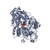
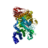
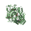
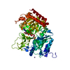
| Title | Structure of the periplasmic binding protein MelB (Atu4661) in complex with melibiose from Agrobacterium fabrum C58 | |||||||||
 Components Components | Periplasmic alpha-galactoside-binding protein | |||||||||
 Keywords Keywords | TRANSPORT PROTEIN / protein transporter | |||||||||
| Function / homology |  Function and homology information Function and homology information | |||||||||
| Biological species |  Rhizobium radiobacter (Agrobacterium genomosp. 4) Rhizobium radiobacter (Agrobacterium genomosp. 4) | |||||||||
| Method |  X-RAY DIFFRACTION / SYNCHROTRON / MOLECULAR REPLACEMENT / Resolution: 1.8 Å X-RAY DIFFRACTION / SYNCHROTRON / MOLECULAR REPLACEMENT / Resolution: 1.8 Å | |||||||||
 Authors Authors | Vigouroux, A. / Morera, S. | |||||||||
| Funding support |  France, 1items France, 1items
| |||||||||
 Citation Citation | Journal: J. Biol. Chem. / Year: 2018 Title: The plant defense signal galactinol is specifically used as a nutrient by the bacterial pathogenAgrobacterium fabrum. Authors: Meyer, T. / Vigouroux, A. / Aumont-Nicaise, M. / Comte, G. / Vial, L. / Lavire, C. / Morera, S. | |||||||||
| History |
|
- Structure visualization
| Structure viewer | Molecule: MolmilJmol/JSmol |
|---|
- Downloads & links
-Download
| PDBx/mmCIF format | 6epz.cif.gz | 1 MB | Display | PDBx/mmCIF format |
|---|---|---|---|---|
| PDB format | pdb6epz.ent.gz | 882.9 KB | Display | PDB format |
| PDBx/mmJSON format | 6epz.json.gz | Tree view | PDBx/mmJSON format | |
| Others |  Other downloads Other downloads |
-Validation report
| Arichive directory | https://data.pdbj.org/pub/pdb/validation_reports/ep/6epzftp://data.pdbj.org/pub/pdb/validation_reports/ep/6epz | HTTPS FTP |
|---|
-Related structure data
| Related structure data |  6epySC  6eq0C 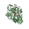 6eq1C 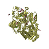 6eq8C S: Starting model for refinement C: citing same article ( |
|---|---|
| Similar structure data |
-Links
 PDBj
PDBj- Assembly
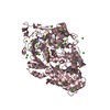
| Deposited unit | 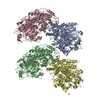
| ||||||||
|---|---|---|---|---|---|---|---|---|---|
| 1 | 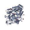
| ||||||||
| 2 | 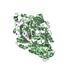
| ||||||||
| 3 | 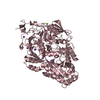
| ||||||||
| 4 | 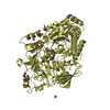
| ||||||||
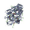
| Unit cell |
| ||||||||
| Components on special symmetry positions |
|
-Components
-Protein / Sugars , 2 types, 8 molecules ADCB
| #1: Protein | Mass: 76359.602 Da / Num. of mol.: 4 Source method: isolated from a genetically manipulated source Source: (gene. exp.) Rhizobium radiobacter (Agrobacterium genomosp. 4)Gene: SY94_4618 / Production host: #2: Polysaccharide | alpha-D-galactopyranose-(1-6)-alpha-D-glucopyranose / alpha-melibiose 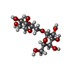  Source method: isolated from a genetically manipulated source Details: oligosaccharide / References: alpha-melibiose |
|---|
-Non-polymers , 6 types, 1416 molecules 


| #3: Chemical | ChemComp-MG /  Mass: 24.305 Da / Num. of mol.: 5 / Source method: obtained synthetically / Formula: Mg Mass: 24.305 Da / Num. of mol.: 5 / Source method: obtained synthetically / Formula: Mg#4: Chemical | ChemComp-EDO /  Mass: 62.068 Da / Num. of mol.: 9 / Source method: obtained synthetically / Formula: C2H6O2 Mass: 62.068 Da / Num. of mol.: 9 / Source method: obtained synthetically / Formula: C2H6O2#5: Chemical | ChemComp-NA /  Mass: 22.990 Da / Num. of mol.: 28 / Source method: obtained synthetically / Formula: Na Mass: 22.990 Da / Num. of mol.: 28 / Source method: obtained synthetically / Formula: Na#6: Chemical | ChemComp-CL / |  Mass: 35.453 Da / Num. of mol.: 1 / Source method: obtained synthetically / Formula: Cl Mass: 35.453 Da / Num. of mol.: 1 / Source method: obtained synthetically / Formula: Cl#7: Chemical | ChemComp-PEG / |  Mass: 106.120 Da / Num. of mol.: 1 / Source method: obtained synthetically / Formula: C4H10O3 Mass: 106.120 Da / Num. of mol.: 1 / Source method: obtained synthetically / Formula: C4H10O3#8: Water | ChemComp-HOH / | Mass: 18.015 Da / Num. of mol.: 1372 / Source method: isolated from a natural source / Formula: H2O |
|---|
-Details
| Has protein modification | Y |
|---|
-Experimental details
-Experiment
| Experiment | Method: X-RAY DIFFRACTION / Number of used crystals: 1 |
|---|
- Sample preparation
| Crystal | Density Matthews: 2.2 Å3/Da / Density % sol: 44.12 % |
|---|---|
| Crystal grow | Temperature: 298 K / Method: vapor diffusion, hanging drop Details: 25% PEG 4000, 0.6 M NaCl, 0.2 M CaCl2 and 0.1 M Mes pH 6.5 |
-Data collection
| Diffraction | Mean temperature: 100 K |
|---|---|
| Diffraction source | Source: SYNCHROTRON / Site: SOLEIL / Beamline: PROXIMA 1 / Wavelength: 0.98 Å |
| Detector | Type: DECTRIS PILATUS3 6M / Detector: PIXEL / Date: May 18, 2017 |
| Radiation | Protocol: SINGLE WAVELENGTH / Monochromatic (M) / Laue (L): M / Scattering type: x-ray |
| Radiation wavelength | Wavelength: 0.98 Å / Relative weight: 1 |
| Reflection | Resolution: 1.8→49 Å / Num. obs: 246319 / % possible obs: 99.4 % / Redundancy: 6.6 % / Biso Wilson estimate: 30.42 Å2 / CC1/2: 0.998 / Rsym value: 0.1 / Net I/σ(I): 11.4 |
| Reflection shell | Resolution: 1.8→1.9 Å / Mean I/σ(I) obs: 1.9 / CC1/2: 0.8 / Rsym value: 0.772 / % possible all: 97.1 |
- Processing
| Software |
| |||||||||||||||||||||||||||||||||||||||||||||||||||||||||||||||||||||||||||||||||||||||||||||||||||||||||||||||||||||||||||||
|---|---|---|---|---|---|---|---|---|---|---|---|---|---|---|---|---|---|---|---|---|---|---|---|---|---|---|---|---|---|---|---|---|---|---|---|---|---|---|---|---|---|---|---|---|---|---|---|---|---|---|---|---|---|---|---|---|---|---|---|---|---|---|---|---|---|---|---|---|---|---|---|---|---|---|---|---|---|---|---|---|---|---|---|---|---|---|---|---|---|---|---|---|---|---|---|---|---|---|---|---|---|---|---|---|---|---|---|---|---|---|---|---|---|---|---|---|---|---|---|---|---|---|---|---|---|---|
| Refinement | Method to determine structure: MOLECULAR REPLACEMENT Starting model: 6EPY Resolution: 1.8→48.85 Å / Cor.coef. Fo:Fc: 0.955 / Cor.coef. Fo:Fc free: 0.948 / Rfactor Rfree error: 0 / SU R Cruickshank DPI: 0.122 / Cross valid method: THROUGHOUT / σ(F): 0 / SU R Blow DPI: 0.122 / SU Rfree Blow DPI: 0.106 / SU Rfree Cruickshank DPI: 0.107
| |||||||||||||||||||||||||||||||||||||||||||||||||||||||||||||||||||||||||||||||||||||||||||||||||||||||||||||||||||||||||||||
| Displacement parameters | Biso mean: 33.82 Å2
| |||||||||||||||||||||||||||||||||||||||||||||||||||||||||||||||||||||||||||||||||||||||||||||||||||||||||||||||||||||||||||||
| Refine analyze | Luzzati coordinate error obs: 0.24 Å | |||||||||||||||||||||||||||||||||||||||||||||||||||||||||||||||||||||||||||||||||||||||||||||||||||||||||||||||||||||||||||||
| Refinement step | Cycle: 1 / Resolution: 1.8→48.85 Å
| |||||||||||||||||||||||||||||||||||||||||||||||||||||||||||||||||||||||||||||||||||||||||||||||||||||||||||||||||||||||||||||
| Refine LS restraints |
| |||||||||||||||||||||||||||||||||||||||||||||||||||||||||||||||||||||||||||||||||||||||||||||||||||||||||||||||||||||||||||||
| LS refinement shell | Resolution: 1.8→1.85 Å / Rfactor Rfree error: 0 / Total num. of bins used: 20
| |||||||||||||||||||||||||||||||||||||||||||||||||||||||||||||||||||||||||||||||||||||||||||||||||||||||||||||||||||||||||||||
| Refinement TLS params. | Method: refined / Refine-ID: X-RAY DIFFRACTION
| |||||||||||||||||||||||||||||||||||||||||||||||||||||||||||||||||||||||||||||||||||||||||||||||||||||||||||||||||||||||||||||
| Refinement TLS group |
|
