 Movie
Movie Controller


+ Open data

- Basic information
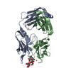
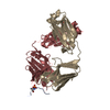
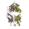
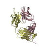
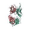
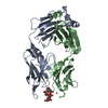
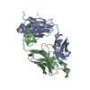
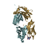
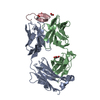
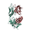
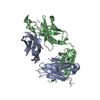
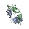
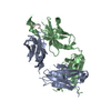
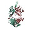
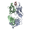
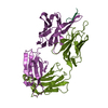
| Entry | Database: PDB / ID: 4m6o | ||||||
|---|---|---|---|---|---|---|---|
| Title | Crystal structure of anti-NGF antibody CNTO7309 | ||||||
 Components Components |
| ||||||
 Keywords Keywords | IMMUNE SYSTEM / immunoglobulin fold / antibody | ||||||
| Function / homology | Immunoglobulins / Immunoglobulin-like / Sandwich / Mainly Beta / NICKEL (II) ION Function and homology information Function and homology information | ||||||
| Biological species |  Homo sapiens (human) Homo sapiens (human) | ||||||
| Method |  X-RAY DIFFRACTION / MOLECULAR REPLACEMENT / Resolution: 2.8 Å X-RAY DIFFRACTION / MOLECULAR REPLACEMENT / Resolution: 2.8 Å | ||||||
 Authors Authors | Teplyakov, A. / Obmolova, G. / Malia, T. / Gilliland, G.L. | ||||||
 Citation Citation | Journal: Proteins / Year: 2014 Title: Antibody modeling assessment II. Structures and models. Authors: Teplyakov, A. / Luo, J. / Obmolova, G. / Malia, T.J. / Sweet, R. / Stanfield, R.L. / Kodangattil, S. / Almagro, J.C. / Gilliland, G.L. | ||||||
| History |
|
- Structure visualization
| Structure viewer | Molecule: MolmilJmol/JSmol |
|---|
- Downloads & links
-Download
| PDBx/mmCIF format | 4m6o.cif.gz | 97.7 KB | Display | PDBx/mmCIF format |
|---|---|---|---|---|
| PDB format | pdb4m6o.ent.gz | 74.8 KB | Display | PDB format |
| PDBx/mmJSON format | 4m6o.json.gz | Tree view | PDBx/mmJSON format | |
| Others |  Other downloads Other downloads |
-Validation report
| Arichive directory | https://data.pdbj.org/pub/pdb/validation_reports/m6/4m6oftp://data.pdbj.org/pub/pdb/validation_reports/m6/4m6o | HTTPS FTP |
|---|
-Related structure data
| Related structure data |  4kmtC  4kq4C 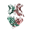 4m6mC  4m7kC  4mauC 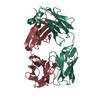 1vgeS 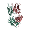 2aabS S: Starting model for refinement C: citing same article ( |
|---|---|
| Similar structure data |


-Links
 PDBj
PDBj

- Assembly
| Deposited unit | 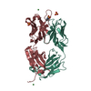
| ||||||||
|---|---|---|---|---|---|---|---|---|---|
| 1 |
| ||||||||
| Unit cell |
| ||||||||
| Components on special symmetry positions |
|
-Components
-Antibody , 2 types, 2 molecules LH
| #1: Antibody | Mass: 23119.627 Da / Num. of mol.: 1 Source method: isolated from a genetically manipulated source Source: (gene. exp.) Homo sapiens (human) / Cell line (production host): HEK 293 / Production host: Homo sapiens (human) |
|---|---|
| #2: Antibody | Mass: 25155.125 Da / Num. of mol.: 1 / Fragment: FD Source method: isolated from a genetically manipulated source Source: (gene. exp.) Homo sapiens (human) / Cell line (production host): HEK 293 / Production host: Homo sapiens (human) |
-Non-polymers , 4 types, 39 molecules 


| #3: Chemical | ChemComp-MES /  Mass: 195.237 Da / Num. of mol.: 1 / Source method: obtained synthetically / Formula: C6H13NO4S / Comment: pH buffer*YM Mass: 195.237 Da / Num. of mol.: 1 / Source method: obtained synthetically / Formula: C6H13NO4S / Comment: pH buffer*YM | ||||
|---|---|---|---|---|---|
| #4: Chemical |  Mass: 96.063 Da / Num. of mol.: 2 / Source method: obtained synthetically / Formula: SO4 Mass: 96.063 Da / Num. of mol.: 2 / Source method: obtained synthetically / Formula: SO4#5: Chemical |  Mass: 58.693 Da / Num. of mol.: 2 / Source method: obtained synthetically / Formula: Ni Mass: 58.693 Da / Num. of mol.: 2 / Source method: obtained synthetically / Formula: Ni#6: Water | ChemComp-HOH / | Mass: 18.015 Da / Num. of mol.: 34 / Source method: isolated from a natural source / Formula: H2O |
-Details
| Has protein modification | Y |
|---|
-Experimental details
-Experiment
| Experiment | Method: X-RAY DIFFRACTION / Number of used crystals: 1 |
|---|
- Sample preparation
| Crystal | Density Matthews: 3.07 Å3/Da / Density % sol: 60 % |
|---|---|
| Crystal grow | Temperature: 293 K / Method: vapor diffusion, sitting drop / pH: 6.5 Details: 0.1 M MES, pH 6.5, 12% PEG20000, VAPOR DIFFUSION, SITTING DROP, temperature 293K |
-Data collection
| Diffraction | Mean temperature: 95 K |
|---|---|
| Diffraction source | Source: ROTATING ANODE / Type: RIGAKU MICROMAX-007 HF / Wavelength: 1.5418 / Wavelength: 1.5418 Å |
| Detector | Type: RIGAKU SATURN 944 / Detector: CCD / Date: Aug 10, 2009 / Details: VARIMAX HF |
| Radiation | Protocol: SINGLE WAVELENGTH / Monochromatic (M) / Laue (L): M / Scattering type: x-ray |
| Radiation wavelength | Wavelength: 1.5418 Å / Relative weight: 1 |
| Reflection | Resolution: 2.8→30 Å / Num. all: 13878 / Num. obs: 13878 / % possible obs: 95.7 % / Observed criterion σ(F): 0 / Observed criterion σ(I): -3 / Redundancy: 3.4 % / Biso Wilson estimate: 35.9 Å2 / Rmerge(I) obs: 0.086 / Net I/σ(I): 14.5 |
| Reflection shell | Resolution: 2.8→2.9 Å / Redundancy: 1.5 % / Rmerge(I) obs: 0.405 / Mean I/σ(I) obs: 1.8 / % possible all: 61.7 |
- Processing
| Software |
| |||||||||||||||||||||||||||||||||||||||||||||
|---|---|---|---|---|---|---|---|---|---|---|---|---|---|---|---|---|---|---|---|---|---|---|---|---|---|---|---|---|---|---|---|---|---|---|---|---|---|---|---|---|---|---|---|---|---|---|
| Refinement | Method to determine structure: MOLECULAR REPLACEMENT Starting model: PDB ENTRIES 1VGE AND 2AAB Resolution: 2.8→15 Å / Cor.coef. Fo:Fc: 0.937 / Cor.coef. Fo:Fc free: 0.874 / SU B: 14.26 / SU ML: 0.267 / Cross valid method: THROUGHOUT / σ(F): 0 / ESU R Free: 0.369 / Stereochemistry target values: Engh & Huber
| |||||||||||||||||||||||||||||||||||||||||||||
| Solvent computation | Ion probe radii: 0.8 Å / Shrinkage radii: 0.8 Å / VDW probe radii: 1.2 Å / Solvent model: BABINET MODEL WITH MASK | |||||||||||||||||||||||||||||||||||||||||||||
| Displacement parameters | Biso mean: 39 Å2 | |||||||||||||||||||||||||||||||||||||||||||||
| Refinement step | Cycle: LAST / Resolution: 2.8→15 Å
| |||||||||||||||||||||||||||||||||||||||||||||
| Refine LS restraints |
| |||||||||||||||||||||||||||||||||||||||||||||
| LS refinement shell | Resolution: 2.815→2.886 Å / Total num. of bins used: 20
|
