 Movie
Movie Controller

[English] 日本語
 Yorodumi
- PDB-4lyp: Crystal Structure of Glycoside Hydrolase Family 5 Mannosidase fro... -
+ Open data

- Basic information
| Entry | Database: PDB / ID: 4lyp | ||||||
|---|---|---|---|---|---|---|---|
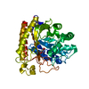
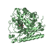
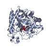
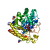
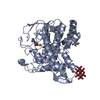
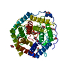
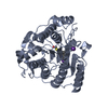
| Title | Crystal Structure of Glycoside Hydrolase Family 5 Mannosidase from Rhizomucor miehei | ||||||
 Components Components | Exo-beta-1,4-mannosidase | ||||||
 Keywords Keywords | HYDROLASE / Tim Barrel / extracellular protein | ||||||
| Function / homology | Glycosidases / TIM Barrel / Alpha-Beta Barrel / Alpha Beta / GUANIDINE Function and homology information Function and homology information | ||||||
| Biological species |  Rhizomucor miehei (fungus) Rhizomucor miehei (fungus) | ||||||
| Method |  X-RAY DIFFRACTION / SYNCHROTRON / MOLECULAR REPLACEMENT / Resolution: 1.28 Å X-RAY DIFFRACTION / SYNCHROTRON / MOLECULAR REPLACEMENT / Resolution: 1.28 Å | ||||||
 Authors Authors | Jiang, Z.Q. / Zhou, P. / Yang, S.Q. / Liu, Y. / Yan, Q.J. | ||||||
 Citation Citation | Journal: Acta Crystallogr.,Sect.D / Year: 2014 Title: Structural insights into the substrate specificity and transglycosylation activity of a fungal glycoside hydrolase family 5 beta-mannosidase. Authors: Zhou, P. / Liu, Y. / Yan, Q. / Chen, Z. / Qin, Z. / Jiang, Z. | ||||||
| History |
|
- Structure visualization
| Structure viewer | Molecule: MolmilJmol/JSmol |
|---|
- Downloads & links
-Download
| PDBx/mmCIF format | 4lyp.cif.gz | 397.5 KB | Display | PDBx/mmCIF format |
|---|---|---|---|---|
| PDB format | pdb4lyp.ent.gz | 325.8 KB | Display | PDB format |
| PDBx/mmJSON format | 4lyp.json.gz | Tree view | PDBx/mmJSON format | |
| Others |  Other downloads Other downloads |
-Validation report
| Arichive directory | https://data.pdbj.org/pub/pdb/validation_reports/ly/4lypftp://data.pdbj.org/pub/pdb/validation_reports/ly/4lyp | HTTPS FTP |
|---|
-Related structure data


-Links
 PDBj
PDBj- Assembly
| Deposited unit | 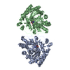
| ||||||||
|---|---|---|---|---|---|---|---|---|---|
| 1 | 
| ||||||||
| 2 | 
| ||||||||
| Unit cell |
|
-Components
| #1: Protein | Mass: 51297.961 Da / Num. of mol.: 2 Source method: isolated from a genetically manipulated source Source: (gene. exp.) Rhizomucor miehei (fungus) / Production host:  #2: Chemical | ChemComp-GAI /   Mass: 59.070 Da / Num. of mol.: 4 / Source method: obtained synthetically / Formula: CH5N3 Mass: 59.070 Da / Num. of mol.: 4 / Source method: obtained synthetically / Formula: CH5N3#3: Chemical |   Mass: 122.143 Da / Num. of mol.: 2 / Source method: obtained synthetically / Formula: C4H12NO3 / Comment: pH buffer*YM Mass: 122.143 Da / Num. of mol.: 2 / Source method: obtained synthetically / Formula: C4H12NO3 / Comment: pH buffer*YM#4: Water | ChemComp-HOH / |  Mass: 18.015 Da / Num. of mol.: 1283 / Source method: isolated from a natural source / Formula: H2O Mass: 18.015 Da / Num. of mol.: 1283 / Source method: isolated from a natural source / Formula: H2OHas protein modification | Y | Sequence details | A SEQUENCE REFERENCE FOR THIS PROTEIN DOES NOT CURRENTLY EXIST. | |
|---|
-Experimental details
-Experiment
| Experiment | Method: X-RAY DIFFRACTION / Number of used crystals: 1 |
|---|
- Sample preparation
| Crystal | Density Matthews: 2.27 Å3/Da / Density % sol: 45.7 % |
|---|---|
| Crystal grow | Temperature: 293 K / Method: vapor diffusion, sitting drop / pH: 7.5 Details: 16% PEG 4000, 0.2 M guanidine hydrochloride, 0.1M HEPES buffer pH 7.5, VAPOR DIFFUSION, SITTING DROP, temperature 293K |
-Data collection
| Diffraction | Mean temperature: 100 K | |||||||||
|---|---|---|---|---|---|---|---|---|---|---|
| Diffraction source | Source: SYNCHROTRON / Site: Photon Factory  / Beamline: AR-NE3A / Wavelength: 0.9794 Å / Beamline: AR-NE3A / Wavelength: 0.9794 Å | |||||||||
| Detector | Type: ADSC QUANTUM 270 / Detector: CCD / Date: May 21, 2012 | |||||||||
| Radiation | Protocol: SINGLE WAVELENGTH / Monochromatic (M) / Laue (L): M / Scattering type: x-ray | |||||||||
| Radiation wavelength | Wavelength: 0.9794 Å / Relative weight: 1 | |||||||||
| Reflection | Resolution: 1.28→86 Å / Num. all: 235043 / Num. obs: 205153 / % possible obs: 87.6 % / Observed criterion σ(F): 2 / Observed criterion σ(I): 2 / Redundancy: 9.9 % / Rmerge(I) obs: 0.052 | |||||||||
| Reflection shell |
|
- Processing
| Software |
| ||||||||||||||||||||||||||||||||||||||||||||||||||||||||||||
|---|---|---|---|---|---|---|---|---|---|---|---|---|---|---|---|---|---|---|---|---|---|---|---|---|---|---|---|---|---|---|---|---|---|---|---|---|---|---|---|---|---|---|---|---|---|---|---|---|---|---|---|---|---|---|---|---|---|---|---|---|---|
| Refinement | Method to determine structure: MOLECULAR REPLACEMENT / Resolution: 1.28→20 Å / Cor.coef. Fo:Fc: 0.976 / Cor.coef. Fo:Fc free: 0.966 / SU B: 0.983 / SU ML: 0.019 / Cross valid method: THROUGHOUT / ESU R: 0.043 / ESU R Free: 0.043 / Stereochemistry target values: MAXIMUM LIKELIHOOD / Details: HYDROGENS HAVE BEEN USED IF PRESENT IN THE INPUT
| ||||||||||||||||||||||||||||||||||||||||||||||||||||||||||||
| Solvent computation | Ion probe radii: 0.8 Å / Shrinkage radii: 0.8 Å / VDW probe radii: 1.2 Å / Solvent model: MASK | ||||||||||||||||||||||||||||||||||||||||||||||||||||||||||||
| Displacement parameters | Biso mean: 14.159 Å2
| ||||||||||||||||||||||||||||||||||||||||||||||||||||||||||||
| Refinement step | Cycle: LAST / Resolution: 1.28→20 Å
| ||||||||||||||||||||||||||||||||||||||||||||||||||||||||||||
| Refine LS restraints |
| ||||||||||||||||||||||||||||||||||||||||||||||||||||||||||||
| LS refinement shell | Resolution: 1.277→1.311 Å / Total num. of bins used: 20
|
