Resolution: 2.6→2.64 Å / Redundancy: 2.7 % / Rmerge(I) obs: 0.33 / Mean I/σ(I) obs: 2.36 / Num. unique all: 1297 / Rsym value: 0.394 / % possible all: 88.3
-
Processing
Software
Name
Version
Classification
HKL-2000
datacollection
REFMAC
5.6.0117
refinement
HKL-2000
datareduction
HKL-2000
datascaling
Refinement
Method to determine structure: MOLECULAR REPLACEMENT / Resolution: 2.57→50 Å / Cor.coef. Fo:Fc: 0.922 / Cor.coef. Fo:Fc free: 0.892 / SU B: 10.827 / SU ML: 0.232 / Cross valid method: THROUGHOUT / ESU R Free: 0.333 / Stereochemistry target values: MAXIMUM LIKELIHOOD / Details: HYDROGENS HAVE BEEN USED IF PRESENT IN THE INPUT
Rfactor
Num. reflection
% reflection
Selection details
Rfree
0.24252
1463
5.2 %
RANDOM
Rwork
0.19544
-
-
-
obs
0.19792
26912
92.4 %
-
Solvent computation
Ion probe radii: 0.8 Å / Shrinkage radii: 0.8 Å / VDW probe radii: 1.2 Å / Solvent model: MASK
 Movie
Movie Controller

 Yorodumi
 Open data
 Basic information
 Components
 Keywords
 Function and homology information
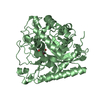
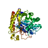
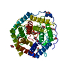
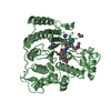
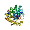
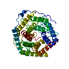
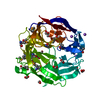
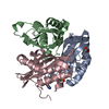
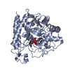
 Rhizomucor miehei (fungus)
 X-RAY DIFFRACTION /
  Authors
 Citation
 Structure visualization
 Downloads & links
 Other downloads


 PDBj
PDBj Assembly


 Mass: 18.015 Da / Num. of mol.: 227 / Source method: isolated from a natural source / Formula: H2O
 Sample preparation
 / Beamline: 3W1A / Wavelength: 1 Å
 Processing