 Movie
Movie Controller

[English] 日本語
 Yorodumi
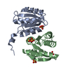
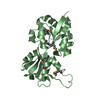
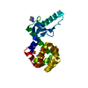
- PDB-4lwm: Crystal structure of methionine sulfoxide reductase U16C/E55D fro... -
+ Open data

- Basic information
| Entry | Database: PDB / ID: 4lwm | |||||||||
|---|---|---|---|---|---|---|---|---|---|---|
| Title | Crystal structure of methionine sulfoxide reductase U16C/E55D from clostridium oremlandii with methionie sulfoxide | |||||||||
 Components Components | Peptide methionine sulfoxide reductase MsrA | |||||||||
 Keywords Keywords | OXIDOREDUCTASE / ALPHA/BETA FOLD / PEPTIDE-METHIONINE (S)-S-OXIDE REDUCTASE | |||||||||
| Function / homology |  Function and homology information Function and homology informationL-methionine (S)-S-oxide reductase activity / peptide-methionine (S)-S-oxide reductase / peptide-methionine (S)-S-oxide reductase activity / protein modification process Similarity search - Function | |||||||||
| Biological species |  Alkaliphilus oremlandii (bacteria) Alkaliphilus oremlandii (bacteria) | |||||||||
| Method |  X-RAY DIFFRACTION / SYNCHROTRON / MOLECULAR REPLACEMENT / Resolution: 1.804 Å X-RAY DIFFRACTION / SYNCHROTRON / MOLECULAR REPLACEMENT / Resolution: 1.804 Å | |||||||||
 Authors Authors | Hwang, K.Y. / Lee, E.H. | |||||||||
 Citation Citation | Journal: Arch.Biochem.Biophys. / Year: 2014 Title: Structural analysis of 1-Cys type selenoprotein methionine sulfoxide reductase A Authors: Lee, E.H. / Kwak, G.H. / Kim, M.J. / Kim, H.Y. / Hwang, K.Y. | |||||||||
| History |
|
- Structure visualization
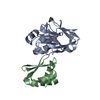
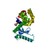
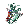
| Structure viewer | Molecule: MolmilJmol/JSmol |
|---|
- Downloads & links
-Download
| PDBx/mmCIF format | 4lwm.cif.gz | 58.9 KB | Display | PDBx/mmCIF format |
|---|---|---|---|---|
| PDB format | pdb4lwm.ent.gz | 41.8 KB | Display | PDB format |
| PDBx/mmJSON format | 4lwm.json.gz | Tree view | PDBx/mmJSON format | |
| Others |  Other downloads Other downloads |
-Validation report
| Summary document | 4lwm_validation.pdf.gz | 452.7 KB | Display | wwPDB validaton report |
|---|---|---|---|---|
| Full document | 4lwm_full_validation.pdf.gz | 453.4 KB | Display | |
| Data in XML | 4lwm_validation.xml.gz | 11.4 KB | Display | |
| Data in CIF | 4lwm_validation.cif.gz | 16 KB | Display | |
| Arichive directory | https://data.pdbj.org/pub/pdb/validation_reports/lw/4lwmftp://data.pdbj.org/pub/pdb/validation_reports/lw/4lwm | HTTPS FTP |
-Related structure data
| Related structure data | 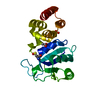 4lwjSC  4lwkC 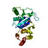 4lwlC  4lwn S: Starting model for refinement C: citing same article ( |
|---|---|
| Similar structure data |


-Links
 PDBj
PDBj- Assembly
| Deposited unit | 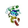
| |||||||||
|---|---|---|---|---|---|---|---|---|---|---|
| 1 |
| |||||||||
| Unit cell |
| |||||||||
| Components on special symmetry positions |
|
-Components
| #1: Protein | Mass: 24786.729 Da / Num. of mol.: 1 / Mutation: (SEC)16C,E55D Source method: isolated from a genetically manipulated source Source: (gene. exp.) Alkaliphilus oremlandii (bacteria) / Strain: OhILAs / Gene: msrA / Plasmid: PET21B / Production host: References: UniProt: A8MI53, peptide-methionine (S)-S-oxide reductase |
|---|---|
| #2: Chemical | ChemComp-MHO / 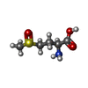  Type: L-peptide linking / Mass: 165.211 Da / Num. of mol.: 1 / Source method: obtained synthetically / Formula: C5H11NO3S Type: L-peptide linking / Mass: 165.211 Da / Num. of mol.: 1 / Source method: obtained synthetically / Formula: C5H11NO3S |
| #3: Chemical | ChemComp-ACT /   Mass: 59.044 Da / Num. of mol.: 1 / Source method: obtained synthetically / Formula: C2H3O2 Mass: 59.044 Da / Num. of mol.: 1 / Source method: obtained synthetically / Formula: C2H3O2 |
| #4: Water | ChemComp-HOH /  Mass: 18.015 Da / Num. of mol.: 157 / Source method: isolated from a natural source / Formula: H2O Mass: 18.015 Da / Num. of mol.: 157 / Source method: isolated from a natural source / Formula: H2O |
-Experimental details
-Experiment
| Experiment | Method: X-RAY DIFFRACTION / Number of used crystals: 1 |
|---|
- Sample preparation
| Crystal | Density Matthews: 2.55 Å3/Da / Density % sol: 51.71 % |
|---|---|
| Crystal grow | Temperature: 293 K / Method: vapor diffusion, hanging drop / pH: 6.5 Details: 0.1M MES, 30%(V/V) PEG 5000, 0.2M AMMONIUM SULFATE, pH 6.5, VAPOR DIFFUSION, HANGING DROP, temperature 293K |
-Data collection
| Diffraction | Mean temperature: 100 K |
|---|---|
| Diffraction source | Source: SYNCHROTRON / Site: PAL/PLS  / Beamline: 6C1 / Wavelength: 1.23985 / Beamline: 6C1 / Wavelength: 1.23985 |
| Detector | Type: ADSC QUANTUM 210 / Detector: CCD / Date: Oct 30, 2010 / Details: SI(111) |
| Radiation | Monochromator: SI(111) / Protocol: SINGLE WAVELENGTH / Monochromatic (M) / Laue (L): M / Scattering type: x-ray |
| Radiation wavelength | Wavelength: 1.23985 Å / Relative weight: 1 |
| Reflection | Resolution: 1.8→30 Å / Num. obs: 22944 / % possible obs: 97.1 % / Observed criterion σ(I): 2 / Biso Wilson estimate: 18.4 Å2 |
| Reflection shell | Resolution: 1.8→1.83 Å / % possible all: 91.5 |
- Processing
| Software |
| |||||||||||||||||||||||||||||||||||||||||||||||||||||||||||||||
|---|---|---|---|---|---|---|---|---|---|---|---|---|---|---|---|---|---|---|---|---|---|---|---|---|---|---|---|---|---|---|---|---|---|---|---|---|---|---|---|---|---|---|---|---|---|---|---|---|---|---|---|---|---|---|---|---|---|---|---|---|---|---|---|---|
| Refinement | Method to determine structure: MOLECULAR REPLACEMENT Starting model: PDB ENTRY 4LWJ Resolution: 1.804→26.464 Å / Occupancy max: 1 / Occupancy min: 0 / FOM work R set: 0.8698 / SU ML: 0.24 / σ(F): 0.37 / Phase error: 20.64 / Stereochemistry target values: ML
| |||||||||||||||||||||||||||||||||||||||||||||||||||||||||||||||
| Solvent computation | Shrinkage radii: 0.9 Å / VDW probe radii: 1.11 Å / Solvent model: FLAT BULK SOLVENT MODEL | |||||||||||||||||||||||||||||||||||||||||||||||||||||||||||||||
| Displacement parameters | Biso max: 69.71 Å2 / Biso mean: 17.1956 Å2 / Biso min: 2.42 Å2 | |||||||||||||||||||||||||||||||||||||||||||||||||||||||||||||||
| Refinement step | Cycle: LAST / Resolution: 1.804→26.464 Å
| |||||||||||||||||||||||||||||||||||||||||||||||||||||||||||||||
| Refine LS restraints |
| |||||||||||||||||||||||||||||||||||||||||||||||||||||||||||||||
| LS refinement shell | Refine-ID: X-RAY DIFFRACTION / Total num. of bins used: 8
|
