 Movie
Movie Controller

[English] 日本語
 Yorodumi
- PDB-1o2f: COMPLEX OF ENZYME IIAGLC AND IIBGLC PHOSPHOCARRIER PROTEIN HPR FR... -
+ Open data

- Basic information
| Entry | Database: PDB / ID: 1o2f | |||||||||
|---|---|---|---|---|---|---|---|---|---|---|
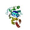
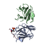
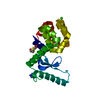
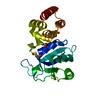
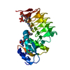
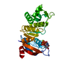
| Title | COMPLEX OF ENZYME IIAGLC AND IIBGLC PHOSPHOCARRIER PROTEIN HPR FROM ESCHERICHIA COLI NMR, RESTRAINED REGULARIZED MEAN STRUCTURE | |||||||||
 Components Components |
| |||||||||
 Keywords Keywords | TRANSFERASE / PHOSPHOTRANSFERASE / KINASE / SUGAR TRANSPORT / COMPLEX (TRANSFERASE-PHOSPHOCARRIER) | |||||||||
| Function / homology |  Function and homology information Function and homology informationprotein-phosphocysteine-glucose phosphotransferase system transporter activity / negative regulation of carbohydrate metabolic process / protein-Npi-phosphohistidine-D-glucose phosphotransferase / regulation of carbohydrate utilization / enzyme IIA-maltose transporter complex / negative regulation of maltose transport / protein-N(PI)-phosphohistidine-carbohydrate phosphotransferase activity / negative regulation of transmembrane transport / D-glucose import across plasma membrane / D-glucose transmembrane transporter activity ...protein-phosphocysteine-glucose phosphotransferase system transporter activity / negative regulation of carbohydrate metabolic process / protein-Npi-phosphohistidine-D-glucose phosphotransferase / regulation of carbohydrate utilization / enzyme IIA-maltose transporter complex / negative regulation of maltose transport / protein-N(PI)-phosphohistidine-carbohydrate phosphotransferase activity / negative regulation of transmembrane transport / D-glucose import across plasma membrane / D-glucose transmembrane transporter activity / D-glucose transmembrane transport / phosphoenolpyruvate-dependent sugar phosphotransferase system / transmembrane transporter complex / kinase activity / regulation of DNA-templated transcription / membrane / metal ion binding / plasma membrane / cytosol Similarity search - Function | |||||||||
| Biological species |  | |||||||||
| Method | SOLUTION NMR / CONJOINED RIGID BODY, TORSION ANGLE DYNAMICS | |||||||||
 Authors Authors | Clore, G.M. / Cai, M. / Williams, D.C. | |||||||||
 Citation Citation | Journal: J.Biol.Chem. / Year: 2003 Title: Solution Structure of the Phosphoryl Transfer Complex between the Signal-transducing Protein IIAGlucose and the Cytoplasmic Domain of the Glucose Transporter IICBGlucose of the Escherichia ...Title: Solution Structure of the Phosphoryl Transfer Complex between the Signal-transducing Protein IIAGlucose and the Cytoplasmic Domain of the Glucose Transporter IICBGlucose of the Escherichia coli Glucose Phosphotransferase System. Authors: Cai, M. / Williams Jr., D.C. / Wang, G. / Lee, B.R. / Peterkofsky, A. / Clore, G.M. | |||||||||
| History |
|
- Structure visualization
| Structure viewer | Molecule: MolmilJmol/JSmol |
|---|
- Downloads & links
-Download
| PDBx/mmCIF format | 1o2f.cif.gz | 229.8 KB | Display | PDBx/mmCIF format |
|---|---|---|---|---|
| PDB format | pdb1o2f.ent.gz | 191.5 KB | Display | PDB format |
| PDBx/mmJSON format | 1o2f.json.gz | Tree view | PDBx/mmJSON format | |
| Others |  Other downloads Other downloads |
-Validation report
| Arichive directory | https://data.pdbj.org/pub/pdb/validation_reports/o2/1o2fftp://data.pdbj.org/pub/pdb/validation_reports/o2/1o2f | HTTPS FTP |
|---|
-Related structure data
| Similar structure data |
|---|


-Links
 PDBj
PDBj- Assembly
| Deposited unit | 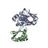
| |||||||||
|---|---|---|---|---|---|---|---|---|---|---|
| 1 |
| |||||||||
| NMR ensembles |
|
-Components
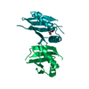
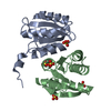
| #1: Protein | Mass: 18141.834 Da / Num. of mol.: 1 Source method: isolated from a genetically manipulated source Source: (gene. exp.) References: UniProt: P69783, protein-Npi-phosphohistidine-sugar phosphotransferase |
|---|---|
| #2: Protein | Mass: 9256.379 Da / Num. of mol.: 1 Source method: isolated from a genetically manipulated source Source: (gene. exp.) References: UniProt: P69786, protein-Npi-phosphohistidine-sugar phosphotransferase |
| #3: Chemical | ChemComp-PO3 /   Mass: 78.972 Da / Num. of mol.: 1 / Source method: obtained synthetically / Formula: PO3 Mass: 78.972 Da / Num. of mol.: 1 / Source method: obtained synthetically / Formula: PO3 |
-Experimental details
-Experiment
| Experiment | Method: SOLUTION NMR | ||||||||||||||||||||||||||||
|---|---|---|---|---|---|---|---|---|---|---|---|---|---|---|---|---|---|---|---|---|---|---|---|---|---|---|---|---|---|
| NMR experiment |
|
- Sample preparation
| Sample conditions | Ionic strength: 10 mM SODIUM PHOSPHATE / pH: 7 / Temperature: 308.00 K |
|---|---|
| Crystal grow | *PLUS Method: other / Details: NMR |
-NMR measurement
| Radiation | Protocol: SINGLE WAVELENGTH / Monochromatic (M) / Laue (L): M | ||||||||||||||||||||||||||||||
|---|---|---|---|---|---|---|---|---|---|---|---|---|---|---|---|---|---|---|---|---|---|---|---|---|---|---|---|---|---|---|---|
| Radiation wavelength | Relative weight: 1 | ||||||||||||||||||||||||||||||
| NMR spectrometer |
|
- Processing
| NMR software | Name:  X-PLOR NIH / Version: (HTTP://NMR.CIT.NIH.GOV/XPLOR_NIH) X-PLOR NIH / Version: (HTTP://NMR.CIT.NIH.GOV/XPLOR_NIH)Developer: SCHWIETERS, KUSZEWSKI, TJANDRA, CLORE J.MAGN.RESON. 160, 66-73 (2003) Classification: refinement | |||||||||||||||
|---|---|---|---|---|---|---|---|---|---|---|---|---|---|---|---|---|
| Refinement | Method: CONJOINED RIGID BODY, TORSION ANGLE DYNAMICS / Software ordinal: 1 Details: THE STRUCTURES WERE CALCULATED BY CONJOINED RIGID BODY/TORSION ANGLE DYNAMICS (SCHWIETERS & CLORE (2001) J.MAGN.RESON 152, 288-302). THE TARGET FUNCTIONS COMPRISES TERMS FOR THE NOE-DERIVED ...Details: THE STRUCTURES WERE CALCULATED BY CONJOINED RIGID BODY/TORSION ANGLE DYNAMICS (SCHWIETERS & CLORE (2001) J.MAGN.RESON 152, 288-302). THE TARGET FUNCTIONS COMPRISES TERMS FOR THE NOE-DERIVED TERMS FOR THE NOE RESTRAINTS (INTRA AND INTERMOLECULAR), THE INTERFACIAL SIDECHAIN TORSION ANGLE RESTRAINTS FOR IIAGLC, THE BACKBONE AND SIDE CHAIN TORSION ANGLE RESTRAINTS FOR IIBGLC, THE DIPOLAR COUPLING RESTRAINTS FOR IIBGL (CLORE ET AL. J.MAGN.RESON. 131, 159-162 (1998); J.MAGN.RESON. 133, 216-221(1998)), THE RADIUS OF GYRATION (KUSZEWSKI ET AL. (1999), A QUARTIC VAN DER WAALS REPULSION TERM (NILGES ET AL. (1988) FEBS LETT. 229, 129- 136), AND A TORSION ANGLE DATABASE POTENTIAL OF MEAN FORCE (CLORE & KUSZEWSKI (2002) J.AM.CHEM.SOC 121, 2337-2338). THE STARTING COORDINATES FOR IIAGLC ARE FROM THE 2.1 ANGSTROM RESOLUTION X-RAY STRUCTURE (WITH PROTONS ADDED) OF E. COLI IIAGLC (MOLECULE 2 OF 2F3G; FEESE ET AL. BIOCHEMISTRY 36, 16087-16096 (1997)). THE BACKBONE COORDINATES AND NON- INTERFACIAL SIDECHAINS OF IIAGLC ARE TREATED AS A RIGID BODY. IN THIS ENTRY THE LAST COLUMN REPRESENTS THE AVERAGE RMS DIFFERENCE BETWEEN THE INDIVIDUAL SIMULATED ANNEALING STRUCTURES AND THE MEAN COORDINATE POSITIONS. IT IS IMPORTANT TO NOTE THAT THE VALUES GIVEN FOR THE BACKBONE ATOMS AND NON-INTERFACIAL SIDECHAINS OF IIAGLC PROVIDE ONLY A MEASURE OF THE PRECISION WITH WHICH THE RELATIVE ORIENTATION OF IIAGLC IN THE COMPLEX HAS BEEN DETERMINED AND DOES NOT TAKE INTO ACCOUNT THE THE ERRORS IN THE X-RAY COORDINATES OF IIAGLC RESIDUE NUMBERING: IIAGLC: 19-168 (RESIDUES 1-18 ARE DISORDERED IN SOLUTION AND NOT VISIBLE IN THE ELECTRON DENSITY MAP OF THE CRYSTAL STRUCTURE OF THE FREE PROTEIN). IIBGLC: 314-390 (CORRESPONDING TO RESIDUES 400-476 OF INTACT IIBCGLC. RESIDUES 301-314 ARE DISORDERED IN SOLUTION. PRO317 HAS BEEN MUTATED TO ALA TO REMOVE HETEROGENEITY ARISING FROM CIS-TRANS PROLINE ISOMERIZATION. PHOSPHATE: RESIDUE 200 EXPERIMENTAL RESTRAINTS: INTRAMOLECULAR INTERPROTON DISTANCE RESTRAINTS: IIBGLC: 987 (189 INTRARESIDUE, 273 SEQUENTIAL, (218 MEDIUM RANGE (1 < |I-J|<=5, 307 LONG RANGE (|I-J>5) IIAGLC INTERFACIAL SIDE CHAINS: 30 INTERMOLECULAR INTERPROTON DISTANCE RESTRAINTS: 113 BACKBONE H-BOND RESTRAINTS FOR IIBGLC (2 PER H-BOND): 72 TORSION ANGLE RESTRAINTS: IIBGLC: 221 IIAGLC INTERFACIAL SIDE CHAINS: 34 RESIDUAL DIPOLAR COUPLINGS FOR IIBGLC: 174 (58 N-H, 58 N-C', 58 HN-C') 13CALPHA/BETA SHIFTS FOR IIBGLC: 138 THREE SETS OF COORDINATES ARE GIVEN: MODEL 3: RESTRAINED REGULARIZED MEAN COORDINATES OF THE UNPHOSPHORYLATED IIAGLC-IIBGLC COMPLEX OVERALL BACKBONE COORDINATE PRECISION (IIAGLC+IIBGLC): 0.31A HEAVY ATOM INTERFACE SIDECHAIN COORDINATE PRECISION (IIAGLC+IIBGLC): 0.67A BACKBONE COORDINATE PRECISION FOR IIBGLC: 0.21 A ALL HEAVY ATOM COORDINATE PRECISION FOR IIBGLC; 0.71 A MODEL 2: RESTRAINED REGULARIZED MEAN COORDINATES FOR THE MODEL OF THE DISSOCIATIVE PHOSPHORYL TRANSITION STATE IIAGLC-IIBGLC. EXPERIMENTAL RESTRAINTS ARE IDENTICAL TO THOSE USED FOR MODEL 3, BUT COVALENT GEOMETRY RESTRAINTS ARE INCLUDED RELATING TO THE PENTACOORDINATE PHOSPHORYL GROUP IN A TRIGONAL BIPYRAMIDAL GEOMETRY. THE STRUCTURE IS DERIVED FROM MODEL 3 BY RESTRAINED CONJOINED TORSION ANGLE/RIGID BODY MINIMIZATION. NO RESTRAINTS WERE EMPLOYED FOR THE NE2(HIS90/IIAGLC)-P AND SG(CYS35/IIBGLC)-P DISTANCES. THERE IS NO CHANGE IN BACKBONE RELATIVE TO MODEL 3 BUT THE NE2(HIS90/IIAGLC- SG(CYS35/IIBGLC) DISTANCE IS REDUCED FROM 5.75 A IN MODEL 3 TO 5.3 A IN MODEL 2. MODEL 1: RESTRAINED REGULARIZED MEAN COORDINATES FOR THE MODEL OF THE ASSOCIATIVE PHOSPHORYL TRANSITION STATE HPR-IIAGLC COMPLEX. CALCULATED LIKE MODEL 2 BUT WITH THE NE2(HIS90/IIAGLC)-P AND SG(CYS35/IIBGLC)-P DISTANCES RESTRAINED TO 2 A. THE STRUCTURE IS DERIVED FROM MODEL 3 BY RESTRAINED CONJOINED TORSION ANGLE/RIGID BODY MINIMIZATION. THE RMS DIFFERENCE BETWEEN THE MEAN STRUCTURES OF THE UNPHOSPHORYLATED COMPLEX (MODEL 3) AND THE TRANSITION STATE COMPLEX (MODEL 1) IS ONLY 0.1 A FOR BACKBONE COORDINATES IMMEDIATELY ADJACENT TO THE ACTIVE SITE HIS AND CYS (RESIDUES 89-91 OF IIAGLC AND 334-336 of IIBGLC). THE REMAINING BACKBONE COORDINATES DO NOT SHIFT. | |||||||||||||||
| NMR ensemble | Conformer selection criteria: REGULARIZED MEAN STRUCTURES / Conformers calculated total number: 60 / Conformers submitted total number: 3 | |||||||||||||||
| Refine LS restraints | *PLUS
|
