 Movie
Movie Controller

[English] 日本語
 Yorodumi
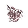
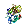
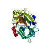
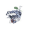
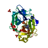
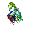
- PDB-4lw3: Crystal structure of the Drosophila beta1,4galactosyltransferase7... -
+ Open data

- Basic information
| Entry | Database: PDB / ID: 4lw3 | ||||||
|---|---|---|---|---|---|---|---|
| Title | Crystal structure of the Drosophila beta1,4galactosyltransferase7 catalytic domain D211N single mutant enzyme complex with manganese and UDP-galactose | ||||||
 Components Components | Beta-4-galactosyltransferase 7 | ||||||
 Keywords Keywords | TRANSFERASE / GT-A glycosyltransferase family / manganese and UDp-galactose / Golgi | ||||||
| Function / homology |  Function and homology information Function and homology informationxylosylprotein 4-beta-galactosyltransferase / xylosylprotein 4-beta-galactosyltransferase activity / glycosaminoglycan-protein linkage region biosynthetic process / Glycosaminoglycan-protein linkage region biosynthesis / proteoglycan biosynthetic process / chondroitin sulfate proteoglycan biosynthetic process / heparan sulfate proteoglycan biosynthetic process / N-glycan processing / carbohydrate metabolic process / Golgi membrane ...xylosylprotein 4-beta-galactosyltransferase / xylosylprotein 4-beta-galactosyltransferase activity / glycosaminoglycan-protein linkage region biosynthetic process / Glycosaminoglycan-protein linkage region biosynthesis / proteoglycan biosynthetic process / chondroitin sulfate proteoglycan biosynthetic process / heparan sulfate proteoglycan biosynthetic process / N-glycan processing / carbohydrate metabolic process / Golgi membrane / nucleotide binding / Golgi apparatus / metal ion binding Similarity search - Function | ||||||
| Biological species |  | ||||||
| Method |  X-RAY DIFFRACTION / MOLECULAR REPLACEMENT / Resolution: 2 Å X-RAY DIFFRACTION / MOLECULAR REPLACEMENT / Resolution: 2 Å | ||||||
 Authors Authors | Ramakrishnan, B. / Qasba, P.K. | ||||||
 Citation Citation | Journal: J.Biol.Chem. / Year: 2013 Title: Crystal Structures of beta-1,4-Galactosyltransferase 7 Enzyme Reveal Conformational Changes and Substrate Binding. Authors: Tsutsui, Y. / Ramakrishnan, B. / Qasba, P.K. | ||||||
| History |
|
- Structure visualization
| Structure viewer | Molecule: MolmilJmol/JSmol |
|---|
- Downloads & links
-Download
| PDBx/mmCIF format | 4lw3.cif.gz | 75.2 KB | Display | PDBx/mmCIF format |
|---|---|---|---|---|
| PDB format | pdb4lw3.ent.gz | 53.3 KB | Display | PDB format |
| PDBx/mmJSON format | 4lw3.json.gz | Tree view | PDBx/mmJSON format | |
| Others |  Other downloads Other downloads |
-Validation report
| Arichive directory | https://data.pdbj.org/pub/pdb/validation_reports/lw/4lw3ftp://data.pdbj.org/pub/pdb/validation_reports/lw/4lw3 | HTTPS FTP |
|---|
-Related structure data
| Related structure data |  4irpC  4irqC 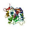 4lw6C 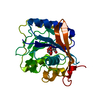 4m4kC  3lw6S S: Starting model for refinement C: citing same article ( |
|---|---|
| Similar structure data |

-Links
 PDBj
PDBj

- Assembly
| Deposited unit | 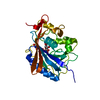
| ||||||||
|---|---|---|---|---|---|---|---|---|---|
| 1 |
| ||||||||
| Unit cell |
| ||||||||
| Components on special symmetry positions |
|
-Components
| #1: Protein | Mass: 33319.305 Da / Num. of mol.: 1 / Fragment: catalytic domain, UNP residues 71-311 / Mutation: D221N Source method: isolated from a genetically manipulated source Details: Protein expressed as inclusion bodies and in vitro refolded Source: (gene. exp.) Gene: 4-galactosyltransferase-7, beta-4GalT7, beta1, beta4GalT7, beta4GalT7-RA, CG11780, Dmel_CG11780 Plasmid: pET23a / Production host:  References: UniProt: Q9VBZ9, Transferases; Glycosyltransferases; Hexosyltransferases, xylosylprotein 4-beta-galactosyltransferase |
|---|---|
| #2: Chemical | ChemComp-MN /   Mass: 54.938 Da / Num. of mol.: 1 / Source method: obtained synthetically / Formula: Mn Mass: 54.938 Da / Num. of mol.: 1 / Source method: obtained synthetically / Formula: Mn |
| #3: Chemical | ChemComp-GDU / 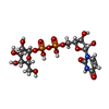  Mass: 566.302 Da / Num. of mol.: 1 Mass: 566.302 Da / Num. of mol.: 1Source method: isolated from a genetically manipulated source Formula: C15H24N2O17P2 |
| #4: Water | ChemComp-HOH /  Mass: 18.015 Da / Num. of mol.: 304 / Source method: isolated from a natural source / Formula: H2O Mass: 18.015 Da / Num. of mol.: 304 / Source method: isolated from a natural source / Formula: H2O |
| Has protein modification | Y |
-Experimental details
-Experiment
| Experiment | Method: X-RAY DIFFRACTION / Number of used crystals: 1 |
|---|
- Sample preparation
| Crystal | Density Matthews: 3.37 Å3/Da / Density % sol: 63.52 % |
|---|---|
| Crystal grow | Temperature: 280 K / Method: vapor diffusion, hanging drop / pH: 8 Details: 100 mM HEPES, 5% PEG 6K, 10% MPD, 2 mM MnCl2, 2 mM UDP-Gal, pH 8.0, VAPOR DIFFUSION, HANGING DROP, temperature 280K |
-Data collection
| Diffraction | Mean temperature: 100 K |
|---|---|
| Diffraction source | Source: ROTATING ANODE / Type: RIGAKU MICROMAX-007 HF / Wavelength: 1.5418 Å |
| Detector | Type: MAR scanner 345 mm plate / Detector: IMAGE PLATE / Date: Jul 17, 2013 / Details: Mirrors |
| Radiation | Monochromator: Graphite / Protocol: SINGLE WAVELENGTH / Monochromatic (M) / Laue (L): M / Scattering type: x-ray |
| Radiation wavelength | Wavelength: 1.5418 Å / Relative weight: 1 |
| Reflection | Resolution: 2→50 Å / Num. obs: 29731 / % possible obs: 97 % / Observed criterion σ(I): 1 / Redundancy: 11.2 % / Rsym value: 0.057 / Net I/σ(I): 36.6 |
| Reflection shell | Resolution: 2→2.07 Å / Redundancy: 6.2 % / Mean I/σ(I) obs: 3.2 / Num. unique all: 2614 / Rsym value: 0.353 / % possible all: 85.4 |
- Processing
| Software |
| ||||||||||||||||||||||||||||||||||||||||||||||||||||||||||||||||||||||||||||||||||||
|---|---|---|---|---|---|---|---|---|---|---|---|---|---|---|---|---|---|---|---|---|---|---|---|---|---|---|---|---|---|---|---|---|---|---|---|---|---|---|---|---|---|---|---|---|---|---|---|---|---|---|---|---|---|---|---|---|---|---|---|---|---|---|---|---|---|---|---|---|---|---|---|---|---|---|---|---|---|---|---|---|---|---|---|---|---|
| Refinement | Method to determine structure: MOLECULAR REPLACEMENT Starting model: PDB entry 3LW6 Resolution: 2→28.913 Å / SU ML: 0.2 / Cross valid method: Throught / σ(F): 1.33 / Phase error: 22.77 / Stereochemistry target values: ML
| ||||||||||||||||||||||||||||||||||||||||||||||||||||||||||||||||||||||||||||||||||||
| Solvent computation | Shrinkage radii: 0.9 Å / VDW probe radii: 1.11 Å / Solvent model: FLAT BULK SOLVENT MODEL | ||||||||||||||||||||||||||||||||||||||||||||||||||||||||||||||||||||||||||||||||||||
| Refinement step | Cycle: LAST / Resolution: 2→28.913 Å
| ||||||||||||||||||||||||||||||||||||||||||||||||||||||||||||||||||||||||||||||||||||
| Refine LS restraints |
| ||||||||||||||||||||||||||||||||||||||||||||||||||||||||||||||||||||||||||||||||||||
| LS refinement shell |
|
