 Movie
Movie Controller

[English] 日本語
 Yorodumi
- PDB-4ltv: Crystal structure of epi-isozizaene synthase from Streptomyces co... -
+ Open data

- Basic information
| Entry | Database: PDB / ID: 4ltv | ||||||
|---|---|---|---|---|---|---|---|
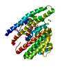
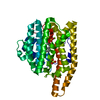
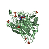
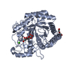
| Title | Crystal structure of epi-isozizaene synthase from Streptomyces coelicolor A3(2) | ||||||
 Components Components | Epi-isozizaene synthase | ||||||
 Keywords Keywords | LYASE / Class I terpene cyclase | ||||||
| Function / homology |  Function and homology information Function and homology informationepi-isozizaene synthase / epi-isozizaene synthase activity / terpene synthase activity / metal ion binding Similarity search - Function | ||||||
| Biological species |  Streptomyces coelicolor (bacteria) Streptomyces coelicolor (bacteria) | ||||||
| Method |  X-RAY DIFFRACTION / SYNCHROTRON / MOLECULAR REPLACEMENT / Resolution: 2.403 Å X-RAY DIFFRACTION / SYNCHROTRON / MOLECULAR REPLACEMENT / Resolution: 2.403 Å | ||||||
 Authors Authors | Li, R. / Chou, W. / Himmelberger, J.A. / Litwin, K. / Harris, G. / Cane, D.E. / Christianson, D.W. | ||||||
 Citation Citation | Journal: Biochemistry / Year: 2014 Title: Reprogramming the Chemodiversity of Terpenoid Cyclization by Remolding the Active Site Contour of epi-Isozizaene Synthase. Authors: Li, R. / Chou, W.K. / Himmelberger, J.A. / Litwin, K.M. / Harris, G.G. / Cane, D.E. / Christianson, D.W. | ||||||
| History |
|
- Structure visualization
| Structure viewer | Molecule: MolmilJmol/JSmol |
|---|
- Downloads & links
-Download
| PDBx/mmCIF format | 4ltv.cif.gz | 79.9 KB | Display | PDBx/mmCIF format |
|---|---|---|---|---|
| PDB format | pdb4ltv.ent.gz | 58.6 KB | Display | PDB format |
| PDBx/mmJSON format | 4ltv.json.gz | Tree view | PDBx/mmJSON format | |
| Others |  Other downloads Other downloads |
-Validation report
| Arichive directory | https://data.pdbj.org/pub/pdb/validation_reports/lt/4ltvftp://data.pdbj.org/pub/pdb/validation_reports/lt/4ltv | HTTPS FTP |
|---|
-Related structure data
| Related structure data | 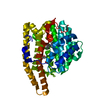 4ltzC  4luuC  4lxwC  4lz0C  4lz3C 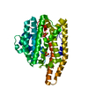 4lzcC  3kb9S S: Starting model for refinement C: citing same article ( |
|---|---|
| Similar structure data |


-Links
 PDBj
PDBj
- Assembly
| Deposited unit | 
| ||||||||
|---|---|---|---|---|---|---|---|---|---|
| 1 |
| ||||||||
| Unit cell |
|
-Components
| #1: Protein | Mass: 43716.012 Da / Num. of mol.: 1 Source method: isolated from a genetically manipulated source Source: (gene. exp.) Streptomyces coelicolor (bacteria) / Strain: A3(2) / Gene: cyc1, SCO5222, SC7E4.19 / Plasmid: pET28a(+) / Production host: | ||
|---|---|---|---|
| #2: Chemical |   Mass: 96.063 Da / Num. of mol.: 2 / Source method: obtained synthetically / Formula: SO4 Mass: 96.063 Da / Num. of mol.: 2 / Source method: obtained synthetically / Formula: SO4#3: Water | ChemComp-HOH / |  Mass: 18.015 Da / Num. of mol.: 53 / Source method: isolated from a natural source / Formula: H2O Mass: 18.015 Da / Num. of mol.: 53 / Source method: isolated from a natural source / Formula: H2O |
-Experimental details
-Experiment
| Experiment | Method: X-RAY DIFFRACTION / Number of used crystals: 1 |
|---|
- Sample preparation
| Crystal | Density Matthews: 2.11 Å3/Da / Density % sol: 41.76 % |
|---|---|
| Crystal grow | Temperature: 298 K / Method: vapor diffusion, sitting drop / pH: 5.5 Details: 4 uL drop of protein solution [8-10 mg/mL EIZS, 20 mM Tris-HCl (pH 7.5), 300 mM NaCl, 10 mM MgCl2, 10% glycerol, 2 mM TCEP, 2 mM sodium pyrophosphate, and 2 mM benzyltriethylammonium ...Details: 4 uL drop of protein solution [8-10 mg/mL EIZS, 20 mM Tris-HCl (pH 7.5), 300 mM NaCl, 10 mM MgCl2, 10% glycerol, 2 mM TCEP, 2 mM sodium pyrophosphate, and 2 mM benzyltriethylammonium chloride] was added to 4 uL of precipitant solution [100 mM Bis-Tris (pH 5.5), 25-28%polyethylene glycol 3350, and 0.2M(NH4)2SO4] and equilibrated against a 1 mL well reservoir of precipitant solution. Crystals appeared within 2-3 days. Then the crystals were soaked in a chelation buffer [5 mM 1,4,7,10-tetraazacyclododecane-1,4,7,10-tetraacetic acid, 15 mM ethylenediaminetetraacetic acid, 100 mM Bis-Tris (pH 5.5), 30% polyethylene glycol 3350, 200 mM (NH4)2SO4, 15% glycerol] for 7 days. , VAPOR DIFFUSION, SITTING DROP, temperature 298K |
-Data collection
| Diffraction | Mean temperature: 100 K |
|---|---|
| Diffraction source | Source: SYNCHROTRON / Site: NSLS  / Beamline: X29A / Wavelength: 1.075 Å / Beamline: X29A / Wavelength: 1.075 Å |
| Detector | Type: ADSC QUANTUM 315 / Detector: CCD / Date: Jan 31, 2012 Details: Monochromator: Double silicon (111) crystal monochromator with cryogenically-cooled first crystal and sagittally-bent second crystal horizontally-focusing at 3.3:1 demagnification. Mirror: ...Details: Monochromator: Double silicon (111) crystal monochromator with cryogenically-cooled first crystal and sagittally-bent second crystal horizontally-focusing at 3.3:1 demagnification. Mirror: Meridionally-bent fused silica mirror with palladium and uncoated stripes vertically-focusing at 6.6:1 demagnification. |
| Radiation | Monochromator: Double silicon (111) crystal monochromator with cryogenically-cooled first crystal and sagittally-bent second crystal horizontally-focusing at 3.3:1 demagnification. Protocol: SINGLE WAVELENGTH / Monochromatic (M) / Laue (L): M / Scattering type: x-ray |
| Radiation wavelength | Wavelength: 1.075 Å / Relative weight: 1 |
| Reflection | Resolution: 2.4→50 Å / Num. obs: 13825 / % possible obs: 95.8 % / Observed criterion σ(F): 0 / Observed criterion σ(I): -3 / Redundancy: 3.1 % / Rmerge(I) obs: 0.091 / Rsym value: 0.091 / Net I/σ(I): 11.912 |
| Reflection shell | Resolution: 2.4→2.49 Å / Redundancy: 2.9 % / Rmerge(I) obs: 0.335 / Mean I/σ(I) obs: 2.423 / Rsym value: 0.335 / % possible all: 94.5 |
- Processing
| Software |
| ||||||||||||||||||||||||||||||||||||||||||||||||||||||||||||||||||||||
|---|---|---|---|---|---|---|---|---|---|---|---|---|---|---|---|---|---|---|---|---|---|---|---|---|---|---|---|---|---|---|---|---|---|---|---|---|---|---|---|---|---|---|---|---|---|---|---|---|---|---|---|---|---|---|---|---|---|---|---|---|---|---|---|---|---|---|---|---|---|---|---|
| Refinement | Method to determine structure: MOLECULAR REPLACEMENT Starting model: PDBID: 3KB9 Resolution: 2.403→41.068 Å / SU ML: 0.32 / σ(F): 0.07 / Phase error: 26.5 / Stereochemistry target values: ML
| ||||||||||||||||||||||||||||||||||||||||||||||||||||||||||||||||||||||
| Solvent computation | Shrinkage radii: 0.9 Å / VDW probe radii: 1.11 Å / Solvent model: FLAT BULK SOLVENT MODEL / Bsol: 61.233 Å2 / ksol: 0.355 e/Å3 | ||||||||||||||||||||||||||||||||||||||||||||||||||||||||||||||||||||||
| Displacement parameters |
| ||||||||||||||||||||||||||||||||||||||||||||||||||||||||||||||||||||||
| Refinement step | Cycle: LAST / Resolution: 2.403→41.068 Å
| ||||||||||||||||||||||||||||||||||||||||||||||||||||||||||||||||||||||
| Refine LS restraints |
| ||||||||||||||||||||||||||||||||||||||||||||||||||||||||||||||||||||||
| LS refinement shell |
|
