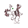
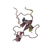
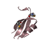
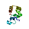
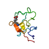
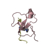
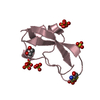
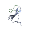
[English] 日本語
Yorodumi
- PDB-4l7x: Crystal structure of the DIDO PHD finger in complex with H3K4me3 -

+
Open data


ID or keywords:

Loading...

-
Basic information

Entry
Database: PDB / ID: 4l7x
TitleCrystal structure of the DIDO PHD finger in complex with H3K4me3
Components
  • Death-inducer obliterator 1
  • Histone H3 peptide
KeywordsCELL CYCLE / GENE REGULATION / mitosis / Chromatin
Function / homology
Function and homology information


Chromatin modifying enzymes / epigenetic regulation of gene expression / Meiotic synapsis / telomere organization / RNA Polymerase I Promoter Opening / Interleukin-7 signaling / Assembly of the ORC complex at the origin of replication / DNA methylation / Condensation of Prophase Chromosomes / HCMV Late Events ...Chromatin modifying enzymes / epigenetic regulation of gene expression / Meiotic synapsis / telomere organization / RNA Polymerase I Promoter Opening / Interleukin-7 signaling / Assembly of the ORC complex at the origin of replication / DNA methylation / Condensation of Prophase Chromosomes / HCMV Late Events / Chromatin modifications during the maternal to zygotic transition (MZT) / ERCC6 (CSB) and EHMT2 (G9a) positively regulate rRNA expression / SIRT1 negatively regulates rRNA expression / PRC2 methylates histones and DNA / Defective pyroptosis / HDACs deacetylate histones / apoptotic signaling pathway / RNA Polymerase I Promoter Escape / Transcriptional regulation by small RNAs / Formation of the beta-catenin:TCF transactivating complex / RUNX1 regulates genes involved in megakaryocyte differentiation and platelet function / Activated PKN1 stimulates transcription of AR (androgen receptor) regulated genes KLK2 and KLK3 / NoRC negatively regulates rRNA expression / B-WICH complex positively regulates rRNA expression / HDMs demethylate histones / spindle / PKMTs methylate histone lysines / RMTs methylate histone arginines / Meiotic recombination / Pre-NOTCH Transcription and Translation / nucleosome assembly / Activation of anterior HOX genes in hindbrain development during early embryogenesis / HCMV Early Events / Transcriptional regulation of granulopoiesis / structural constituent of chromatin / nucleosome / gene expression / RUNX1 regulates transcription of genes involved in differentiation of HSCs / chromatin organization / Factors involved in megakaryocyte development and platelet production / HATs acetylate histones / Senescence-Associated Secretory Phenotype (SASP) / Oxidative Stress Induced Senescence / Estrogen-dependent gene expression / cadherin binding / Amyloid fiber formation / protein heterodimerization activity / DNA-templated transcription / protein-containing complex / DNA binding / RNA binding / extracellular exosome / extracellular region / nucleoplasm / membrane / metal ion binding / nucleus / cytoplasm
Similarity search - Function
Death-inducer obliterator 1 / Spen paralogue and orthologue SPOC, C-terminal / SPOC domain / Transcription factor S-II (TFIIS), central domain / Domain in the central regions of transcription elongation factor S-II (and elsewhere) / Transcription elongation factor S-II, central domain / Transcription elongation factor S-II, central domain superfamily / TFIIS central domain profile. / Zinc/RING finger domain, C3HC4 (zinc finger) / Herpes Virus-1 ...Death-inducer obliterator 1 / Spen paralogue and orthologue SPOC, C-terminal / SPOC domain / Transcription factor S-II (TFIIS), central domain / Domain in the central regions of transcription elongation factor S-II (and elsewhere) / Transcription elongation factor S-II, central domain / Transcription elongation factor S-II, central domain superfamily / TFIIS central domain profile. / Zinc/RING finger domain, C3HC4 (zinc finger) / Herpes Virus-1 / Zinc finger, PHD-type, conserved site / PHD-finger / Zinc finger PHD-type signature. / Zinc finger PHD-type profile. / Zinc finger, PHD-finger / Histone H3 signature 1. / Zinc finger, PHD-type / PHD zinc finger / Histone H3 signature 2. / Histone H3 / Histone H3/CENP-A / Histone H2A/H2B/H3 / Core histone H2A/H2B/H3/H4 / Zinc finger, FYVE/PHD-type / Histone-fold / Zinc finger, RING/FYVE/PHD-type / 2-Layer Sandwich / Alpha Beta
Similarity search - Domain/homology
Histone H3.1 / Death-inducer obliterator 1
Similarity search - Component
Biological speciesHomo sapiens (human)
MethodX-RAY DIFFRACTION / SYNCHROTRON / SAD / Resolution: 1.35 Å
AuthorsTong, Q. / Gatchalian, J. / Kutateladze, T.G.
CitationJournal: Cell Rep / Year: 2013
Title: Dido3 PHD Modulates Cell Differentiation and Division.
Authors: Gatchalian, J. / Futterer, A. / Rothbart, S.B. / Tong, Q. / Rincon-Arano, H. / Sanchez de Diego, A. / Groudine, M. / Strahl, B.D. / Martinez-A, C. / van Wely, K.H. / Kutateladze, T.G.
History
DepositionJun 14, 2013Deposition site: RCSB / Processing site: RCSB
Revision 1.0Jul 24, 2013Provider: repository / Type: Initial release

-
Structure visualization

Structure viewerMolecule:
MolmilJmol/JSmol

Downloads & links

-
Assembly

Deposited unit
A: Death-inducer obliterator 1
U: Histone H3 peptide
hetero molecules


Theoretical massNumber of molelcules
Total (without water)8,7624
Polymers8,6312
Non-polymers1312
Water1,33374
1


  • Idetical with deposited unit
  • defined by author&software
TypeNameSymmetry operationNumber
identity operation1_555x,y,z1
Buried area1240 Å2
ΔGint-4 kcal/mol
Surface area4300 Å2
MethodPISA
Unit cell
Length a, b, c (Å)40.820, 40.820, 80.620
Angle α, β, γ (deg.)90.00, 90.00, 90.00
Int Tables number92
Space group name H-MP41212

-
Components

#1: Protein Death-inducer obliterator 1 / DIO-1 / hDido1 / Death-associated transcription factor 1 / DATF-1


Mass: 7280.341 Da / Num. of mol.: 1 / Fragment: PHD-type zinc finger domain residues 266-325
Source method: isolated from a genetically manipulated source
Source: (gene. exp.) Homo sapiens (human) / Gene: DIDO1, C20orf158, DATF1, KIAA0333 / Production host: Escherichia coli (E. coli) / References: UniProt: Q9BTC0
#2: Protein/peptide Histone H3 peptide / H3K4me3 peptide


Mass: 1350.568 Da / Num. of mol.: 1 / Source method: obtained synthetically / Details: Peptide was synthesized. / Source: (synth.) Homo sapiens (human) / References: UniProt: P68431*PLUS
#3: Chemical ChemComp-ZN / ZINC ION


Mass: 65.409 Da / Num. of mol.: 2 / Source method: obtained synthetically / Formula: Zn
#4: Water ChemComp-HOH / water / Water


Mass: 18.015 Da / Num. of mol.: 74 / Source method: isolated from a natural source / Formula: H2O

-
Experimental details

-
Experiment

ExperimentMethod: X-RAY DIFFRACTION / Number of used crystals: 1

-
Sample preparation

CrystalDensity Matthews: 1.95 Å3/Da / Density % sol: 36.78 %
Crystal growTemperature: 291 K / Method: vapor diffusion, hanging drop / pH: 8
Details: 0.1M HEPES pH8, 30% PEG6000, VAPOR DIFFUSION, HANGING DROP, temperature 291K

-
Data collection

DiffractionMean temperature: 100 K
Diffraction sourceSource: SYNCHROTRON / Site: NSLS / Beamline: X25 / Wavelength: 1 Å
DetectorType: PSI PILATUS 6M / Detector: PIXEL / Date: Feb 26, 2013
RadiationMonochromator: Si-111 double crystal / Protocol: SINGLE WAVELENGTH / Monochromatic (M) / Laue (L): M / Scattering type: x-ray
Radiation wavelengthWavelength: 1 Å / Relative weight: 1
ReflectionResolution: 1.35→28.7 Å / Num. obs: 28593 / % possible obs: 100 % / Observed criterion σ(F): 2 / Observed criterion σ(I): 2
Reflection shellResolution: 1.35→1.4 Å / % possible all: 100

-
Processing

Software
NameVersionClassification
PHENIXmodel building
PHENIX(phenix.refine: 1.8.1_1168)refinement
MOSFLMdata reduction
SCALAdata scaling
PHENIXphasing
RefinementMethod to determine structure: SAD / Resolution: 1.35→28.682 Å / SU ML: 0.07 / σ(F): 1.35 / Phase error: 12.82 / Stereochemistry target values: ML
RfactorNum. reflection% reflectionSelection details
Rfree0.1422 1425 4.98 %RANDOM
Rwork0.1324 ---
all0.14 28593 --
obs0.1329 28593 99.96 %-
Solvent computationShrinkage radii: 0.9 Å / VDW probe radii: 1.11 Å / Solvent model: FLAT BULK SOLVENT MODEL
Refinement stepCycle: LAST / Resolution: 1.35→28.682 Å
ProteinNucleic acidLigandSolventTotal
Num. atoms518 0 2 74 594
Refine LS restraints
Refine-IDTypeDev idealNumber
X-RAY DIFFRACTIONf_bond_d0.007528
X-RAY DIFFRACTIONf_angle_d1.169713
X-RAY DIFFRACTIONf_dihedral_angle_d15.32201
X-RAY DIFFRACTIONf_chiral_restr0.07672
X-RAY DIFFRACTIONf_plane_restr0.00796
LS refinement shell
Resolution (Å)Rfactor RfreeNum. reflection RfreeRfactor RworkNum. reflection RworkRefine-ID% reflection obs (%)
1.35-1.39830.18111460.14692711X-RAY DIFFRACTION100
1.3983-1.45420.16821160.12662758X-RAY DIFFRACTION100
1.4542-1.52040.16581620.11112704X-RAY DIFFRACTION100
1.5204-1.60060.15861490.10612696X-RAY DIFFRACTION100
1.6006-1.70090.11931700.10222700X-RAY DIFFRACTION100
1.7009-1.83220.12391430.10782693X-RAY DIFFRACTION100
1.8322-2.01650.14111280.11212734X-RAY DIFFRACTION100
2.0165-2.30820.13071360.1192731X-RAY DIFFRACTION100
2.3082-2.90760.1351360.1482722X-RAY DIFFRACTION100
2.9076-28.68850.14751390.15042719X-RAY DIFFRACTION100

+
About Yorodumi

-
News

-
Feb 9, 2022. New format data for meta-information of EMDB entries

New format data for meta-information of EMDB entries

  • Version 3 of the EMDB header file is now the official format.
  • The previous official version 1.9 will be removed from the archive.

Related info.:EMDB header

External links:wwPDB to switch to version 3 of the EMDB data model

-
Aug 12, 2020. Covid-19 info

Covid-19 info

URL: https://pdbj.org/emnavi/covid19.php

New page: Covid-19 featured information page in EM Navigator.

Related info.:Covid-19 info / Mar 5, 2020. Novel coronavirus structure data

+
Mar 5, 2020. Novel coronavirus structure data

Novel coronavirus structure data

Related info.:Yorodumi Speices / Aug 12, 2020. Covid-19 info

External links:COVID-19 featured content - PDBj / Molecule of the Month (242):Coronavirus Proteases

+
Jan 31, 2019. EMDB accession codes are about to change! (news from PDBe EMDB page)

EMDB accession codes are about to change! (news from PDBe EMDB page)

  • The allocation of 4 digits for EMDB accession codes will soon come to an end. Whilst these codes will remain in use, new EMDB accession codes will include an additional digit and will expand incrementally as the available range of codes is exhausted. The current 4-digit format prefixed with “EMD-” (i.e. EMD-XXXX) will advance to a 5-digit format (i.e. EMD-XXXXX), and so on. It is currently estimated that the 4-digit codes will be depleted around Spring 2019, at which point the 5-digit format will come into force.
  • The EM Navigator/Yorodumi systems omit the EMD- prefix.

Related info.:Q: What is EMD? / ID/Accession-code notation in Yorodumi/EM Navigator

External links:EMDB Accession Codes are Changing Soon! / Contact to PDBj

+
Jul 12, 2017. Major update of PDB

Major update of PDB

  • wwPDB released updated PDB data conforming to the new PDBx/mmCIF dictionary.
  • This is a major update changing the version number from 4 to 5, and with Remediation, in which all the entries are updated.
  • In this update, many items about electron microscopy experimental information are reorganized (e.g. em_software).
  • Now, EM Navigator and Yorodumi are based on the updated data.

External links:wwPDB Remediation / Enriched Model Files Conforming to OneDep Data Standards Now Available in the PDB FTP Archive

-
Yorodumi

Thousand views of thousand structures

  • Yorodumi is a browser for structure data from EMDB, PDB, SASBDB, etc.
  • This page is also the successor to EM Navigator detail page, and also detail information page/front-end page for Omokage search.
  • The word "yorodu" (or yorozu) is an old Japanese word meaning "ten thousand". "mi" (miru) is to see.

Related info.:EMDB / PDB / SASBDB / Comparison of 3 databanks / Yorodumi Search / Aug 31, 2016. New EM Navigator & Yorodumi / Yorodumi Papers / Jmol/JSmol / Function and homology information / Changes in new EM Navigator and Yorodumi

Read more